Chemisorption Analysis Essentials: Unlocking Catalyst Surface Properties for Advanced Research
This comprehensive guide explores the fundamentals, methodologies, and critical applications of chemisorption for catalyst surface analysis, tailored for researchers and drug development professionals.
Chemisorption Analysis Essentials: Unlocking Catalyst Surface Properties for Advanced Research
Abstract
This comprehensive guide explores the fundamentals, methodologies, and critical applications of chemisorption for catalyst surface analysis, tailored for researchers and drug development professionals. It covers foundational principles of selective gas adsorption, practical techniques like pulse chemisorption and TPD/TPR, troubleshooting common experimental challenges, and validation through complementary surface science tools. The article provides a systematic framework for extracting accurate surface area, active site density, dispersion, and metal particle size data, essential for rational catalyst design and optimization in biomedical and chemical synthesis applications.
Core Principles of Chemisorption: Understanding the Molecule-Surface Bond
1. Introduction & Thesis Context
This whitepaper provides a definitive technical guide to chemisorption, framed within a critical thesis on Fundamentals of chemisorption for catalyst surface analysis research. Accurate distinction between chemisorption and physisorption is the cornerstone of characterizing active sites, determining dispersion, and rationalizing activity and selectivity in heterogeneous catalysis. For researchers in catalysis, materials science, and drug development (where adsorption phenomena underpin drug delivery and sensor platforms), a precise understanding of these mechanisms is non-negotiable.
2. Fundamental Distinctions: Mechanism and Energetics
The primary distinction lies in the nature of the adsorbate-substrate bond.
- Chemisorption involves the formation of a chemical bond (covalent, ionic, or strong polar) via significant electron rearrangement between the adsorbate and the surface atoms. This process is site-specific, often irreversible, and characterized by a high enthalpy change.
- Physical Adsorption (Physisorption) arises from weak, non-specific van der Waals forces or dipole interactions. No electron transfer or chemical bond formation occurs; it is reversible and multi-layered.
The quantitative differences are summarized in Table 1.
Table 1: Quantitative Comparison of Physisorption and Chemisorption
| Property | Physisorption | Chemisorption |
|---|---|---|
| Binding Forces | van der Waals, dipole-dipole | Chemical bonds (covalent, ionic) |
| Enthalpy Change (ΔH) | Low (≈ 5 – 40 kJ/mol) | High (≈ 40 – 800 kJ/mol) |
| Activation Energy | Usually none (non-activated) | Often significant (activated process) |
| Specificity | Non-specific | Highly specific to surface sites/geometry |
| Temperature Range | Occurs near adsorbate boiling point | Occurs at higher temperatures |
| Reversibility | Fully reversible | Often irreversible or requires high T to desorb |
| Layer Formation | Multi-layer | Strictly mono-layer |
| Electron Transfer | No orbital overlap, no electron transfer | Significant orbital overlap, possible electron transfer |
3. Experimental Protocols for Distinction
3.1. Temperature-Programmed Desorption (TPD) TPD is the premier technique for differentiating adsorption mechanisms by probing the strength and distribution of adsorbate binding.
- Protocol: 1) Clean the catalyst surface under inert gas flow at elevated temperature. 2) Cool to adsorption temperature (e.g., 100 K for physisorption studies, 300 K for chemisorption). 3) Expose to a precise dose of probe gas (e.g., CO, NH₃, H₂). 4) Purge with inert gas (He, Ar) to remove physisorbed species. 5) Heat the sample linearly (e.g., 10 K/min) under inert flow. 6) Monitor desorbing species with a mass spectrometer or TCD.
- Interpretation: Physisorbed species desorb at low temperatures (e.g., <150 K for N₂). Chemisorbed species desorb in distinct peaks at higher temperatures, with peak temperature correlating to bond strength. Multiple peaks indicate heterogeneous surface sites.
3.2. Adsorption Isotherm Analysis (BET vs. Chemisorption) Volumetric or gravimetric isotherm measurements can isolate the chemisorbed monolayer.
- Protocol for Metal Dispersion (H₂ or CO Pulse Chemisorption): 1) Pre-treat catalyst (reduction in H₂, then evacuation). 2) Cool to analysis temperature (e.g., 35°C for H₂ on Pt). 3) Inject calibrated pulses of probe gas into a flowing inert carrier gas passing over the sample. 4) Monitor uptake via TCD until consecutive pulses give identical signals, indicating saturation of active sites. 5) Calculate active metal surface area assuming a stoichiometry (e.g., H:Ptₛᵤʳfᴀᴄᴇ = 1:1, CO:Ptₛᵤʀfᴀᴄᴇ = 1:1).
- Interpretation: The total uptake at monolayer saturation, distinct from the multi-layer formation in BET physisorption, quantifies the number of active sites.
4. Visualization of Key Concepts
Diagram 1: Key distinctions between Physisorption and Chemisorption (83 characters)
Diagram 2: Temperature-Programmed Desorption (TPD) workflow (59 characters)
5. The Scientist's Toolkit: Key Research Reagent Solutions & Materials
Table 2: Essential Materials for Chemisorption Experiments
| Item | Typical Example | Function / Rationale |
|---|---|---|
| Probe Gases | 5-10% H₂/Ar, 5-10% CO/He, O₂, NH₃, NO | Chemisorb selectively to specific sites (H₂ on metals, NH₃ on acids) for site counting and strength measurement. |
| Inert Carrier Gases | Ultra-high purity (UHP) Argon, Helium (99.999%) | Provide inert atmosphere for purging and thermal conductivity detection (TCD). He is preferred for TCD sensitivity. |
| Reference Material | Certified SiO₂ or Al₂O₅ powder, traceable metal foils | Calibrate instrument dead volume and validate gas uptake quantification. |
| Catalyst Reduction Gas | UHP Hydrogen (99.999%) | Pre-treatment gas to reduce metal oxides to their active metallic state prior to chemisorption analysis. |
| Calibration Loops | Fixed-volume stainless steel loops (e.g., 0.5, 1.0 cm³) | Deliver precise, reproducible doses of probe gas for pulse chemisorption measurements. |
| Molecular Sieves / Gas Purifiers | 5Å, 13X sieves; oxygen/moisture traps | Remove trace H₂O and O₂ from carrier and probe gases to prevent oxidation of sensitive catalysts during analysis. |
| Thermocouples | K-type (NiCr-NiAl), shielded | Accurate, real-time temperature measurement and control during TPD ramps and isothermal steps. |
The Role of Selective Probe Molecules (H2, CO, O2) in Active Site Titration
Within the broader thesis on the Fundamentals of Chemisorption for Catalyst Surface Analysis Research, the quantification of catalytically active sites—active site titration—is a cornerstone. This guide details the use of selective probe molecules (H₂, CO, O₂) to titrate specific active sites on heterogeneous catalyst surfaces. By exploiting the distinct chemisorptive properties of these gases, researchers can move beyond total surface area measurements to quantify the number and sometimes the nature of sites responsible for catalytic activity.
Theoretical Foundations of Selective Chemisorption
Chemisorption involves the formation of strong, specific chemical bonds between the probe molecule and surface atoms. The selectivity arises from the electronic and geometric structure of the surface sites:
- H₂: Dissociatively chemisorbs on transition metals (e.g., Pt, Pd, Ni) and can titrate metallic sites. It is often used for metals supported on oxides.
- CO: Chemisorbs on both metallic and oxidized sites, but its bonding mode (linear, bridged, carbonyl) provides structural information via infrared spectroscopy. It is highly selective for metal sites.
- O₂: Reacts irreversibly with reduced metal surfaces (e.g., Cu, Ag, Group VIII metals) via dissociative adsorption or oxidation, useful for titrating surface metal atoms.
Table 1: Chemisorptive Properties of Probe Molecules
| Probe Molecule | Typical Target Sites | Stoichiometry (Molecule:Site) | Common Detection Methods | Temperature Range |
|---|---|---|---|---|
| Hydrogen (H₂) | Reduced metal atoms (Pt, Pd, Ni, Co) | 1 H₂ : 2 M (dissociative) | Volumetric, TPD, H₂-O₂ titration | 25-100°C |
| Carbon Monoxide (CO) | Metal atoms (reduced or partially oxidized) | 1 CO : 1 M (linear) or 2 CO : 1 M (bridged) | Volumetric, FTIR, TPD | -80 to 50°C |
| Oxygen (O₂) | Reduced metal atoms (Cu, Ag, Fe, Group VIII) | 1 O₂ : 2 M (dissociative) | Volumetric, pulse titration, TPO | 25-400°C |
Table 2: Comparison of Titration Methodologies
| Parameter | Static Volumetric | Pulse Chemisorption | Titration of Pre-adsorbed Species |
|---|---|---|---|
| Principle | Measures pressure change in a known volume. | Injects pulses into carrier over catalyst until saturation. | Uses a second gas to titrate a pre-adsorbed layer (e.g., H₂-O₂). |
| Accuracy | High (Primary method) | Good (Relative method) | High (for specific systems) |
| Speed | Slow (Equilibrium needed) | Fast | Moderate |
| Data Output | Isotherm, uptake at saturation | Uptake at saturation | Stoichiometric consumption |
| Best For | H₂, O₂, detailed isotherms | Routine quality control, CO | Dispersion of supported metals |
Detailed Experimental Protocols
Protocol: Static Volumetric Titration of Metal Sites using H₂
Objective: To determine the number of reduced surface metal atoms on a Pt/Al₂O₃ catalyst.
- Sample Preparation: ~0.2 g of catalyst is loaded into a quartz sample cell. The sample is degassed under vacuum (<10⁻⁵ mbar) at 120°C for 1 hour to remove physisorbed species.
- Reduction Pretreatment: The sample is reduced in situ under flowing H₂ (50 mL/min) at 400°C for 2 hours, followed by evacuation at 400°C for 1 hour to create a clean, reduced surface.
- Isotherm Measurement: The sample is cooled to the titration temperature (35°C). Precise doses of high-purity H₂ are introduced into the calibrated volume manifold. After each dose, the system is allowed to reach equilibrium (2-5 min), and the pressure is recorded.
- Data Analysis: The total chemisorbed volume (at STP) is determined from the plateau of the adsorption isotherm. Using the stoichiometry (1 H₂ molecule dissociates on 2 Pt atoms), the number of surface Pt atoms and thus the metal dispersion (%) is calculated.
Protocol: Pulse Chemisorption of CO for Metal Dispersion
Objective: Rapid determination of exposed metal sites on a Pd/SiO₂ catalyst.
- Activation: The catalyst sample is reduced in situ in a U-tube reactor under H₂ flow at 300°C, then flushed with inert He at 350°C, and cooled to 35°C in He.
- Pulse Titration: A calibrated pulse loop (e.g., 50 µL) is filled with 10% CO/He mixture. Repeated pulses are injected into the He carrier gas flowing over the catalyst and into a thermal conductivity detector (TCD).
- Endpoint Detection: Pulses are injected until the detector signal shows no further adsorption (consecutive peak areas are constant). The number of pulses consumed is recorded.
- Calculation: The total CO uptake is calculated from pulse size and number of pulses consumed. Assuming a stoichiometry (e.g., CO:Pd = 1:1 for linear adsorption), the metal surface area is derived.
Protocol: O₂ Titration for Copper Surface Area
Objective: To measure the metallic copper surface area in a reduced Cu/ZnO/Al₂O₃ catalyst.
- Sample Reduction: The catalyst is reduced in situ under H₂ at 250°C and evacuated.
- N₂O Reactive Chemisorption: At 60°C, N₂O is introduced (or pulsed) to selectively oxidize surface Cu atoms to Cu₂O: 2 Cu(s) + N₂O → Cu₂O + N₂.
- H₂ Reduction of Surface Oxide: The formed Cu₂O layer is then titrated by temperature-programmed reduction (TPR) with H₂, or by pulsed H₂ at 150°C.
- Calculation: The H₂ consumed in step 3 corresponds to the reduction of oxygen atoms chemisorbed in step 2. Knowing that one O atom bonds to two surface Cu atoms allows for the calculation of the copper metal surface area.
Visualizations
Active Site Titration General Workflow
Selective Probe Molecules & Their Analytical Output
The Scientist's Toolkit: Key Research Reagent Solutions & Materials
| Item | Function & Specification |
|---|---|
| High-Purity Probe Gases | H₂ (99.999%), CO (99.97%), O₂ (99.999%). Essential for quantitative uptake without interference from impurities. |
| Inert Carrier/Blanket Gas | Ultra-high purity He (99.999%) or Ar. Used for purging, as a carrier in pulse flow systems, and for dead volume calibration. |
| Reducing Gas Mixture | 5-10% H₂ in Ar (or N₂). Used for in situ catalyst activation (reduction pretreatment) prior to titration. |
| Nitrous Oxide (N₂O) | For the selective oxidation of surface atoms (e.g., Cu, Co) in indirect oxygen chemisorption methods. |
| Calibrated Pulse Loop | A gas sampling valve with a fixed volume loop (e.g., 50 µL to 1 mL) for precise delivery of gas pulses in flow systems. |
| Reference Material | Certified reference catalysts (e.g., EuroPt-1) with known metal dispersion for validating experimental protocols and setups. |
| Chemisorption Analyzer | Automated instrument combining a vacuum system, calibrated volumes, pressure transducers, and a TCD for static volumetric or pulse chemisorption. |
| In Situ Cell | A reactor cell compatible with both high-temperature pretreatment and low-temperature adsorption, often with IR-transparent windows for FTIR studies. |
Within the context of a thesis on Fundamentals of Chemisorption for Catalyst Surface Analysis Research, understanding the key thermodynamic and kinetic parameters is paramount. Chemisorption, the formation of strong, localized chemical bonds between adsorbate molecules and a catalyst surface, is the critical first step in most heterogeneous catalytic reactions. Two parameters are central to characterizing this process: the Heat of Adsorption (ΔHads) and the Activation Energy (Ea). The heat of adsorption governs the stability of the adsorbed state and influences surface coverage, while the activation energy dictates the rate at which adsorption (or the subsequent surface reaction) occurs. This guide provides an in-depth technical analysis of these parameters, their determination, and their interpretation for researchers and scientists in catalysis and related fields.
Fundamental Concepts
Heat of Adsorption (ΔH_ads): This is the enthalpy change released (exothermic, negative value) or absorbed (endothermic, positive value) when a molecule is chemisorbed onto a surface. For chemisorption, it is typically exothermic and large in magnitude (40-800 kJ/mol), reflecting the strength of the chemical bond formed. It is a thermodynamic parameter that determines the equilibrium coverage of adsorbates at a given temperature and pressure (via the adsorption isotherm). The differential heat of adsorption often varies with surface coverage due to surface heterogeneity and adsorbate-adsorbate interactions.
Activation Energy (Ea): In the context of chemisorption, this refers to the energy barrier that must be overcome for the adsorption process to proceed. It is the minimum kinetic energy required for an incoming molecule to form a chemical bond with the surface. For non-activated adsorption, Ea ≈ 0. For activated adsorption, E_a > 0, meaning the rate of adsorption increases significantly with temperature. This is a kinetic parameter derived from the temperature dependence of the adsorption rate constant (via the Arrhenius equation).
Experimental Methodologies for Determination
Calorimetry for Heat of Adsorption
Protocol: Direct measurement of heat flow during gas adsorption.
- A clean, degassed catalyst sample is placed in a microcalorimeter cell under high vacuum.
- The sample is maintained at a constant temperature (e.g., 300 K).
- Small, precise doses of the adsorbate gas (e.g., CO, H₂) are introduced sequentially to the sample.
- The heat released upon each dose is measured by sensitive thermopiles or heat-flow sensors.
- The corresponding amount adsorbed is measured volumetrically or gravimetrically.
- The differential heat of adsorption is calculated as the heat released per mole of gas adsorbed for each dose, plotted as a function of surface coverage.
Temperature-Programmed Desorption (TPD) for Ea and ΔHads
Protocol: Analysis of desorption kinetics as a function of temperature.
- The catalyst surface is saturated with the adsorbate at a low temperature.
- The system is evacuated to remove physisorbed and gas-phase species.
- The temperature is increased linearly (β = dT/dt, e.g., 10 K/min) under a flow of inert gas or vacuum.
- The desorption rate (pressure or mass signal) is monitored as a function of temperature, yielding TPD spectra (peaks).
- Activation Energy for Desorption (Ed): Estimated using the Redhead or Chan-Aris-Weinberg methods (for first-order desorption): Ed ≈ RTp * ln(νTp / β) - 3.64RTp, where Tp is the peak maximum temperature and ν is the pre-exponential factor (often assumed ~10¹³ s⁻¹).
- Heat of Adsorption: For non-dissociative, immobile adsorption, Ed ≈ -ΔHads, as the activation energy for adsorption (E_a) is often small.
Adsorption Kinetic Studies for Activation Energy of Adsorption
Protocol: Measuring uptake rates at different temperatures.
- A fresh catalyst sample is cleaned and brought to a specific temperature (T1).
- A step change in adsorbate pressure is introduced.
- The increase in sample mass (gravimetric) or decrease in pressure (volumetric) is recorded as a function of time until equilibrium.
- The initial rate of adsorption is extracted from the steepest slope of the uptake curve.
- Steps 1-4 are repeated at several different temperatures (T1, T2, T3...).
- The initial rates (or rate constants) are plotted in an Arrhenius plot: ln(rate) vs. 1/T.
- The slope of the linear fit equals -E_a/R, yielding the activation energy for adsorption.
Data and Comparative Analysis
Table 1: Typical Ranges for Key Parameters in Chemisorption Systems
| Adsorbate | Catalyst Surface | Heat of Adsorption (ΔH_ads) [kJ/mol] | Activation Energy for Adsorption (E_a) [kJ/mol] | Common Measurement Technique |
|---|---|---|---|---|
| Carbon Monoxide (CO) | Pt(111) | 140 - 160 | ~0 (non-activated) | Microcalorimetry, TPD |
| Hydrogen (H₂) | Ni(110) | 95 - 110 | 5 - 15 (activated dissoc.) | TPD, Adsorption Kinetics |
| Nitrogen (N₂) | Fe (Haber-Bosch) | ~200 | 20 - 50 (highly activated) | Calorimetry, TPD |
| Oxygen (O₂) | Ag(111) | 200 - 300 | Low for molecular, high for dissoc. | TPD, XPS |
Table 2: Core Equations for Parameter Calculation
| Parameter | Fundamental Equation | Key Variables | Application Note |
|---|---|---|---|
| Differential Heat of Adsorption | qdiff = (δQ/δn)T,A | Q=Heat, n=moles adsorbed | Measured directly via calorimetry. |
| Integrated Heat of Adsorption | ΔHads = (1/ntotal) ∫ q_diff dn | n_total=total uptake | Average bond strength. |
| Activation Energy (Arrhenius) | k = A exp(-E_a/RT) | k=rate constant, A=pre-exp. factor | Derived from kinetic uptake data. |
| Activation Energy for Desorption (Redhead) | Ed ≈ RTp [ln(νT_p/β) - 3.64] | T_p=TPD peak temp, β=heating rate | Assumes first-order desorption, ν≈10¹³ s⁻¹. |
Visualizing Relationships and Workflows
Title: Energy Pathway for Activated Chemisorption
Title: Microcalorimetry Experimental Workflow
The Scientist's Toolkit: Key Research Reagents & Materials
Table 3: Essential Materials for Chemisorption Parameter Studies
| Item / Reagent Solution | Primary Function | Technical Note |
|---|---|---|
| Single Crystal or Well-Defined Catalyst | Provides a uniform, clean surface for fundamental measurement. | Pt(111), SiO₂-supported Ni nanoparticles. Essential for avoiding data convolution from heterogeneity. |
| Ultra-High Purity (UHP) Gases | Source of adsorbate (CO, H₂, O₂) and inert purge (He, Ar). | 99.999% purity minimizes contamination and side reactions on the surface. |
| Microcalorimeter (e.g., BT2.15) | Directly measures minute heats of adsorption with high sensitivity. | Coupled with volumetric system for simultaneous uptake measurement. |
| Volumetric (Manometric) Setup | Precisely measures the quantity of gas adsorbed onto the catalyst. | Consists of calibrated volumes, precision pressure transducers, and UHV valves. |
| Temperature-Programmed Desorption (TPD) System | Heats sample linearly to probe adsorption strength/kinetics. | Includes mass spectrometer (QMS) for monitoring desorbing species. |
| Ultra-High Vacuum (UHV) System | Creates a clean environment (<10⁻⁹ mbar) for surface preparation and study. | Prevents contamination of the catalyst surface prior to and during experiment. |
| Calibration Gas Mixtures | For calibrating mass spectrometers in TPD or residual gas analyzers. | Known concentrations of adsorbate in inert gas (e.g., 1% CO in He). |
| Ion Sputtering Gun / Annealing Apparatus | For cleaning and reconstructing single crystal surfaces. | Removes impurities via Ar+ bombardment and orders surface via high-T annealing. |
The Langmuir Isotherm and Its Assumptions for Monolayer Coverage
Within the fundamental study of chemisorption for catalyst surface analysis research, the Langmuir isotherm stands as a cornerstone theoretical model. It provides the essential framework for quantifying the adsorption of gas molecules onto a solid surface, a process critical to heterogeneous catalysis, sensor design, and drug delivery system development. This whitepaper elucidates the Langmuir model, its underlying assumptions, experimental validation protocols, and its indispensable role in deriving key surface parameters such as active site density and adsorption enthalpy, which are pivotal for rational catalyst design and analysis.
Core Theory and Assumptions
The Langmuir isotherm describes a dynamic equilibrium between gas-phase molecules and adsorbed molecules on a surface. Its derivation rests on four critical assumptions:
- Monolayer Coverage: Adsorption is limited to a single, complete layer of molecules at the surface (a monolayer).
- Uniform Surface: All adsorption sites are energetically identical and equivalent.
- No Interaction: There is no interaction (attractive or repulsive) between adsorbed molecules.
- Dynamic Equilibrium: The process is reversible, with equal rates of adsorption and desorption at equilibrium.
The fundamental equation is: [ \theta = \frac{KP}{1 + KP} ] where (\theta) is the fractional surface coverage, (P) is the gas pressure, and (K) is the adsorption equilibrium constant, related to the Gibbs free energy of adsorption.
Key Quantitative Relationships and Data
The linearized form of the Langmuir isotherm is used for experimental data fitting: [ \frac{P}{n} = \frac{P}{nm} + \frac{1}{K nm} ] where (n) is the amount adsorbed at pressure (P), and (n_m) is the monolayer capacity.
Table 1: Key Parameters Derived from the Langmuir Isotherm
| Parameter | Symbol | Derivation | Significance in Catalyst Analysis |
|---|---|---|---|
| Monolayer Capacity | (n_m) | Intercept & slope of linear plot | Total number of active adsorption sites (site density). |
| Adsorption Constant | (K) | Slope & intercept of linear plot | Affinity of adsorbate for surface; related to adsorption strength. |
| Surface Coverage | (\theta) | (n / n_m) | Fraction of active sites occupied under given conditions. |
| Gibbs Free Energy | (\Delta G_{ads}^\circ) | (-RT \ln(K)) | Thermodynamic spontaneity of the adsorption process. |
Experimental Protocols for Validation
Volumetric (Manometric) Adsorption Measurement
This is the standard method for determining gas adsorption isotherms.
Protocol:
- Degassing: The solid catalyst sample is placed in a sealed, temperature-controlled cell and evacuated under high vacuum (<10⁻⁵ mbar) at elevated temperature (e.g., 300°C for 12 hours) to remove pre-adsorbed contaminants.
- System Calibration: The void volume of the sample cell is precisely determined using helium expansion.
- Dosing: A known amount of adsorbate gas (e.g., N₂, CO, H₂) is introduced into the sample cell from a reference volume.
- Equilibration: Pressure is monitored until equilibrium is reached (typically 5-30 minutes per point).
- Calculation: The amount adsorbed, (n), is calculated from the pressure drop using the ideal gas law and known volumes.
- Isotherm Construction: Steps 3-5 are repeated across a range of pressures at a constant temperature to generate an adsorption isotherm.
Data Analysis for Langmuir Parameters
- Plotting: Transform the (n) vs. (P) data into a linear Langmuir plot: (P/n) vs. (P).
- Linear Regression: Perform a least-squares linear fit. A high correlation coefficient (R² > 0.99) suggests adherence to the Langmuir model.
- Parameter Extraction:
- Slope = (1 / nm)
- Intercept = (1 / (K nm))
- Validation: Replot the data as (\theta) vs. (P) using calculated (n_m) and (K) to visually assess the fit to the theoretical curve.
Visualizing the Langmuir Adsorption Process
The Scientist's Toolkit: Key Research Reagents & Materials
Table 2: Essential Materials for Langmuir Isotherm Experiments
| Item | Function & Specification |
|---|---|
| High-Surface-Area Catalyst | The substrate for adsorption (e.g., Pt/Al₂O₃, Zeolite, Activated Carbon). Must be fully characterized (BET surface area, pore volume). |
| Ultra-High Purity (UHP) Gases | Adsorbates like N₂ (77 K for physisorption), CO, H₂, O₂ (for chemisorption). Purity >99.999% to prevent surface poisoning. |
| Volumetric Adsorption Analyzer | Instrument (e.g., Micromeritics, Quantachrome) with precise pressure transducers and temperature-controlled bath. |
| High-Vacuum System | Turbo-molecular or diffusion pumps capable of achieving <10⁻⁷ mbar for sample degassing. |
| Sample Cell with Heater | For in situ degassing of the catalyst at defined temperatures (up to 500°C). |
| Calibration Gas (Helium) | Used for dead-volume determination of the sample cell. |
| Liquid Coolant (LN₂, LAr) | For maintaining constant cryogenic temperature (e.g., 77 K) during physisorption measurements. |
| Data Analysis Software | For non-linear curve fitting and linear regression of adsorption data (e.g., Origin, custom scripts in Python/R). |
Within the broader thesis on the Fundamentals of Chemisorption for Catalyst Surface Analysis Research, establishing a quantitative link between chemisorption capacity and intrinsic catalyst properties is paramount. Chemisorption, the formation of strong, specific chemical bonds between gas-phase probe molecules and surface atoms, provides the primary experimental window into quantifying active sites. This guide details how chemisorption measurements are rigorously connected to the core metrics of active site density, metal dispersion, and average particle size, forming the bedrock of heterogeneous catalyst characterization.
Core Theoretical Framework
The total volume of gas chemisorbed (at standard temperature and pressure, STP) is directly proportional to the number of surface atoms, provided the chemisorption stoichiometry is known.
Active Sites (A): Total number of surface metal atoms accessible to the probe molecule. ( A = (V{ads} \cdot NA) / Vm ) where ( V{ads} ) is the chemisorbed gas volume (STP), ( NA ) is Avogadro's number, and ( Vm ) is the molar gas volume at STP (22,414 cm³/mol).
Metal Dispersion (D): The fraction of total metal atoms exposed on the surface. ( D = (Number\ of\ surface\ metal\ atoms) / (Total\ number\ of\ metal\ atoms) ) From chemisorption: ( D = (V_{ads} \cdot S \cdot M) / (f \cdot m \cdot \rho) ) where ( S ) is the chemisorption stoichiometry (probe molecules per surface atom), ( M ) is the atomic weight of the metal, ( f ) is the weight fraction of metal in the sample, ( m ) is the sample mass, and ( \rho ) is the metal density.
Average Particle Size (d): Assuming a regular particle geometry (typically spherical or cubic), the volume-to-surface-area ratio yields the particle size. For spherical particles of uniform size: ( d (nm) = (k \cdot \phi) / D ) where ( k ) is a geometric factor (e.g., 6000 for Pd, Pt assuming spherical particles and H₂ chemisorption with S=1), ( \phi ) is the volume-to-surface area shape factor (6 for spheres), and ( D ) is dispersion (as a decimal).
Table 1: Chemisorption Stoichiometries and Calculation Factors for Common Probe Gases
| Probe Gas | Target Metal | Typical Stoichiometry (S) | Common Assumption | Key Consideration |
|---|---|---|---|---|
| Hydrogen (H₂) | Pt, Pd, Ni, Co | 1 H₂ per 1 surface atom (H/Mₛ = 1) | Dissociative adsorption on metals. | Susceptible to hydrogen spillover to support. |
| Carbon Monoxide (CO) | Pt, Pd, Rh, Ru | 1 CO per 1 surface atom (CO/Mₛ = 1) | Linear adsorption. | Can also bridge (CO/Mₛ = 0.5) on some metals/sites. |
| Oxygen (O₂) | Ag, Cu, Co | 2 O atoms per 1 surface atom (O/Mₛ = 2) | Dissociative adsorption. | Reactive, may form subsurface/bulk oxide. |
| Nitric Oxide (NO) | Pt, Pd, Co | Complex (often assumed 1:1) | Multiple bonding modes. | Requires careful calibration and analysis. |
| Table 2: Calculated Particle Size vs. Dispersion for Spherical Platinum Nanoparticles (H₂ Chemisorption, S=1, k≈6000) | ||||
| Dispersion, D (%) | Number of Atoms per Particle (approx.) | Average Particle Diameter, d (nm) | ||
| :--- | :--- | :--- | ||
| 100 | ~300 | 1.0 | ||
| 60 | ~1,700 | 1.7 | ||
| 40 | ~4,500 | 2.5 | ||
| 20 | ~25,000 | 5.0 | ||
| 10 | ~180,000 | 10.0 | ||
| 5 | ~1,500,000 | 20.0 |
Experimental Protocols
Protocol 1: Static Volumetric (Manometric) Chemisorption Analysis
- Principle: Measures pressure change in a calibrated volume upon gas exposure to the catalyst.
- Procedure:
- Sample Preparation (~100-500 mg): Load catalyst into a quartz cell. Attach to analysis port.
- In-situ Pretreatment: Evacuate (<10⁻⁵ mbar). Heat in flowing gas (e.g., H₂ at 350°C for reduction, O₂ for oxidation). Cool to analysis temperature (often 35°C) under vacuum.
- Calibration: Precisely measure sample loop and analysis cell volumes using non-adsorbing helium.
- Isotherm Measurement: Admit small, sequential doses of probe gas (H₂, CO) into the sample cell. Allow equilibrium after each dose (<0.1 mbar/min change).
- Data Reduction: For each dose, calculate the amount adsorbed using the real gas law (e.g., Peng-Robinson). Plot total adsorption vs. equilibrium pressure.
- Extrapolation: For H₂ on metals, extrapolate the linear, high-pressure region of the total isotherm to zero pressure to determine the strong chemisorption volume, discounting weak physisorption.
Protocol 2: Dynamic Pulse Chemisorption
- Principle: Pulses of probe gas are passed over the catalyst in an inert carrier; unadsorbed gas is quantified.
- Procedure:
- Sample Preparation & Pretreatment: As in Protocol 1, but often in a U-shaped tube.
- Carrier Flow: Establish steady flow of inert gas (Ar, He) through the sample.
- Pulsing: Inject repeated, calibrated pulses of probe gas (e.g., 5% CO/He) into the carrier stream upstream of the catalyst.
- Detection: Use a downstream thermal conductivity detector (TCD) to measure the signal for each pulse. Pulses are adsorbed until the surface is saturated, after which a full pulse elutes.
- Calculation: Sum the gas volume not detected (difference between pulse size and eluted peak area) across all pulses until saturation to find total chemisorbed volume.
Diagrams
Title: Linking Chemisorption Data to Catalyst Metrics
Title: Static Volumetric Chemisorption Workflow
The Scientist's Toolkit: Key Research Reagent Solutions & Materials
Table 3: Essential Materials for Chemisorption Experiments
| Item | Function & Specification | Critical Notes |
|---|---|---|
| High-Purity Probe Gases | Source of adsorbate molecules for site counting. H₂ (99.999%), CO (99.97%), O₂ (99.999%), mixed with inert balance (He/Ar). | Must be ultra-pure to prevent surface contamination by CO, H₂O, or hydrocarbons. Use in-line purifiers. |
| Inert Carrier/Calibration Gas | He (99.999%) for system calibration and as carrier in pulse flow experiments. | High thermal conductivity for TCD detection. Must be purified. |
| Catalyst Sample Tube | Quartz or glass U-tube/reactor for holding catalyst during analysis. | Must withstand high-temperature pretreatment and vacuum. Quartz is preferred for reduction steps. |
| Reference Catalyst | Certified metal on support (e.g., 5% Pt/Al₂O³, 2% Pd/SiO₂) with known dispersion. | Used for validating instrument performance and experimental protocol accuracy. |
| Temperature Program Controller | Controls furnace for precise in-situ pretreatment (oxidation, reduction, desorption). | Enables reproducible thermal history, critical for cleaning and activating surfaces. |
| High-Vacuum System | Combination of turbomolecular and diaphragm backing pumps. | Achieves <10⁻⁶ mbar for sample degassing and preventing contamination in static systems. |
| Pressure Transducers | Capacitance manometers for accurate pressure measurement across wide ranges (e.g., 0-1000 mbar). | Essential for precise dose quantification in volumetric systems. Requires regular calibration. |
| Thermal Conductivity Detector (TCD) | Detects changes in gas composition in the effluent stream during pulse chemisorption. | Calibrated response allows quantification of unadsorbed gas in each pulse. |
Practical Chemisorption Techniques: From Theory to Laboratory Data
Within the broader thesis on Fundamentals of Chemisorption for Catalyst Surface Analysis Research, understanding the historical and methodological evolution of gas adsorption techniques is paramount. Static volumetric analysis, a foundational manometric technique, served as the direct precursor to the modern Brunauer-Emmett-Teller (BET) method. This guide details its core principles and protocols, focusing on its critical role in quantifying monolayer chemisorption capacity—a key parameter in determining the number of active sites on a catalytic surface.
Core Principles and Distinction from Physisorption
Static volumetric analysis measures the amount of a probe gas (e.g., H₂, CO, O₂) adsorbed onto a solid catalyst surface at equilibrium conditions. Unlike the BET method, which leverages multilayer physisorption (typically of N₂ at 77 K) to determine total surface area, static volumetric analysis for chemisorption uses specific gases that form a stoichiometric, single chemical bond (monolayer) with surface sites at elevated temperatures. The key distinction is the irreversibility of adsorption under experimental conditions; strongly chemisorbed species are not removed by simple evacuation, allowing for the selective measurement of active sites.
Modern Experimental Protocol: Hydrogen Chemisorption on a Metal Catalyst
The following is a detailed methodology for a standard hydrogen pulse chemisorption experiment, a derivative of static volumetric analysis commonly used in contemporary research.
Pre-Treatment (Activation)
- Weighing & Loading: Approximately 0.1-0.5 g of catalyst is accurately weighed and loaded into a U-shaped quartz sample cell.
- Dehydration: The sample is heated under a flow of inert gas (e.g., He, Ar) to 150°C (or as specified) for 1 hour to remove physisorbed water.
- Reduction/Oxidation: The sample is then subjected to a temperature-programmed reduction (TPR) or oxidation (TPO) using a specific gas mixture (e.g., 5% H₂/Ar for reduction) at a defined ramp rate (e.g., 10°C/min) to a target temperature (e.g., 500°C for metal oxide reduction). This step is held for 1-2 hours to generate clean, accessible metal surfaces.
- Evacuation: The system is evacuated to high vacuum (<10⁻⁵ Torr) and the sample is cooled to the analysis temperature (often 35°C or 300 K for H₂ chemisorption).
Saturation Chemisorption Measurement
- Dosing: Small, calibrated pulses of pure hydrogen are introduced into the carrier gas stream (He) flowing over the catalyst.
- Detection: A thermal conductivity detector (TCD) downstream monitors the effluent. Initially, each pulse is completely adsorbed by the catalyst, yielding no H₂ signal.
- Saturation Point: Pulses continue until the detector signal indicates breakthrough, i.e., H₂ is no longer being adsorbed. Subsequent pulses show identical peak areas.
- Calculation: The total volume of H₂ consumed prior to breakthrough represents the volume required for monolayer saturation ((V_m)). This is converted to moles of H₂ adsorbed.
Data Analysis
The metal dispersion ((D)), particle size ((d)), and active surface area are calculated assuming a stoichiometry (H:Metal). For example, for platinum, H:Pt = 1:1 is often assumed. [ D (\%) = \frac{(Number\ of\ surface\ metal\ atoms)}{(Total\ number\ of\ metal\ atoms)} \times 100 ] [ d (nm) = \frac{k}{(Metal\ Surface\ Area\ per\ gram\ of\ metal)} ] where (k) is a geometric factor dependent on the metal and particle shape.
Table 1: Key Parameters for Common Chemisorption Probe Gases
| Probe Gas | Typical Analysis Temperature | Common Catalyst Target | Assumed Stoichiometry (Gas:Metal) | Primary Information Obtained |
|---|---|---|---|---|
| Hydrogen (H₂) | 35°C (300 K) | Pt, Pd, Ni, Co, Ru | 1:1 or 2:1* | Metal Dispersion, Active Surface Area |
| Carbon Monoxide (CO) | 35°C (300 K) | Pt, Pd, Rh, Ru | 1:1 (linear) or 2:1 (bridged) | Metal Dispersion, Surface Coordination |
| Oxygen (O₂) | -78°C (195 K) or 35°C | Ag, Cu, Base Metals | O:Metal varies | Active Metal Area, Uptake Capacity |
| Nitric Oxide (NO) | 35°C (300 K) | Cu-Zeolites, Transition Metals | NO:Active Site varies | Active Site Count for SCR reactions |
*H:Pt=1:1 is standard; H:Ni=1:1 is common, but H:Ru=2:1 may be used.
Table 2: Comparison: Static Volumetric Chemisorption vs. BET Physisorption
| Feature | Static Volumetric Chemisorption | BET Physisorption (N₂ at 77 K) |
|---|---|---|
| Primary Goal | Quantify active surface sites (chemically specific) | Determine total surface area & pore texture |
| Probe Gas | Reactive (H₂, CO, O₂) | Inert (N₂, Ar, Kr) |
| Temperature | Often elevated (35-400°C) | Cryogenic (77 K for N₂) |
| Nature of Bond | Strong, irreversible (chemical) | Weak, reversible (physical, multilayer) |
| Key Assumption | Stoichiometry of adsorption (e.g., H:Pt=1:1) | Cross-sectional area of adsorbate molecule |
| Typical Output | Metal dispersion, active site density | Total SSA (m²/g), pore volume & distribution |
The Scientist's Toolkit: Key Research Reagent Solutions & Materials
Table 3: Essential Materials for Static Volumetric Chemisorption Analysis
| Item | Function & Specification |
|---|---|
| High-Purity Probe Gases | H₂ (99.999%), CO (99.97%), O₂ (99.995%). Essential for accurate uptake measurement without interference from impurities. |
| Ultra-High Purity Carrier Gas | He or Ar (99.9999%). Used for purging, sample activation, and as a carrier in pulse chemisorption. |
| Quartz Sample Tube/U-Cell | Inert, high-temperature resistant vessel to hold catalyst sample during pre-treatment and analysis. |
| Calibrated Dosage Loop | Precision volume loop (e.g., 0.1-1.0 cm³) for introducing repeatable pulses of probe gas in pulse chemisorption. |
| Thermal Conductivity Detector (TCD) | Detects the concentration of the probe gas in the carrier stream, indicating adsorption saturation. |
| High-Vacuum System | Combination of turbomolecular and diaphragm pumps to achieve ultimate pressure <10⁻⁵ Torr for sample degassing. |
| Catalyst Reference Standard | Certified material (e.g., alumina-supported Pt) with known metal dispersion for system calibration and validation. |
| Temperature-Controlled Furnace | Provides precise, programmable heating up to 1000°C for sample activation (reduction/oxidation). |
Visualization of Experimental Workflow
Diagram 1: Chemisorption Analysis Workflow (Static Volumetric/Pulse)
Diagram 2: Distinguishing Chemisorption from Physisorption in Isotherms
Within the broader thesis on the Fundamentals of Chemisorption for Catalyst Surface Analysis Research, Dynamic Pulse Chemisorption (DPC) stands as a pivotal quantitative technique. Unlike static volumetric methods, DPC probes the accessible metal surface area, active site concentration, and dispersion of supported catalysts by introducing precisely controlled pulses of a probe gas (e.g., H₂, CO, O₂) into a flowing carrier stream. This guide details the core principles, setup, calibration, and a rigorous protocol for generating reproducible and meaningful data fundamental to catalyst characterization in both academic and industrial research, including pharmaceutical catalyst development for hydrogenation or oxidation processes.
Core Principles & Setup
A DPC system consists of several key modules:
- Gas Delivery System: Mass Flow Controllers (MFCs) for carrier (typically Ar or He) and probe gases.
- Pulse Injection System: A calibrated sampling loop (e.g., 0.1-1.0 mL) integrated into a multi-port valve.
- Reactor: A quartz or stainless-steel U-tube where the solid catalyst sample is held.
- Conditioning Unit: An oven for in-situ temperature-programmed pretreatment (e.g., reduction, oxidation).
- Detection System: A Thermal Conductivity Detector (TCD) is most common, measuring the concentration of unadsorbed probe gas in the carrier stream.
- Data Acquisition: Software to record and integrate the TCD signal peaks.
Diagram 1: Schematic of a Dynamic Pulse Chemisorption Setup (75 chars)
System Calibration
Accurate quantification requires calibration of both the pulse volume and the TCD response.
3.1. Loop Volume Calibration The volume of the sample loop is determined by pulsing a non-adsorbing gas (e.g., Ar) through a bypass line into a calibrated volume (e.g., a bubble flowmeter) at room temperature and pressure.
Protocol:
- Connect a bubble flowmeter to the system outlet.
- With the valve in LOAD position, flush the loop with Ar for 2 minutes.
- Switch the valve to INJECT and simultaneously start the flowmeter timer.
- Measure the volume of gas displaced over 5-10 pulses. Average the volume per pulse.
- Calculate the loop volume at STP using the ideal gas law.
3.2. TCD Response Calibration (K-Factor) The TCD response factor (K, in μV·s·μmol⁻¹) relates the integrated peak area to the molar amount of gas.
Protocol:
- Install an empty reactor tube or one filled with an inert material (quartz wool).
- Set the carrier and probe gas flows to experimental values (e.g., 30 mL/min Ar, 0.5 mL H₂ pulses).
- At the analysis temperature, inject a series of 5-10 identical probe gas pulses. The detector should see the full, unadsorbed pulse.
- Precisely integrate the area (A) for each peak.
- The known amount of gas in each pulse (n, in μmol) is calculated from the calibrated loop volume (P, T).
- The K-factor is: K = A / n. Use the average K from all pulses.
Table 1: Typical Calibration Data Summary
| Calibration Type | Parameter | Typical Value Range | Key Equation |
|---|---|---|---|
| Loop Volume | Measured Volume (Room T, P) | 0.25 - 1.0 mL | VSTP = Vmeas * (Pamb/PSTP) * (TSTP/Tamb) |
| TCD Response | K-Factor (K) | 0.5 - 5.0 μV·s·μmol⁻¹ | K = (Peak Area [μV·s]) / (Moles Injected [μmol]) |
| System Reproducibility | Peak Area RSD | < 2% | RSD = (Std. Dev. / Mean) * 100% |
Step-by-Step Experimental Protocol
Step 1: Sample Preparation & Loading. Weigh an appropriate mass of catalyst (typically 50-200 mg) to give a measurable uptake. Load it into the reactor between quartz wool plugs.
Step 2: In-Situ Pretreatment. This is critical to clean the surface. A common reduction protocol:
- Heat from room temperature to 150°C at 10°C/min in Ar flow (30 mL/min). Hold for 30 min.
- Switch to 5% H₂/Ar (30 mL/min).
- Heat to the target reduction temperature (e.g., 500°C) at 5-10°C/min. Hold for 1-2 hours.
- Cool in flowing H₂/Ar to the analysis temperature (often 35-50°C).
- Flush with pure Ar for 30-60 minutes to remove dissolved hydrogen and establish a stable baseline.
Step 3: Pulse Chemisorption Analysis.
- Set carrier gas flow (e.g., Ar at 30 mL/min). Stabilize TCD baseline at analysis temperature.
- Configure the automated pulse sequence: Injection time (e.g., 30 s), interval between pulses (e.g., 2-3 min).
- Begin pulsing the probe gas (e.g., 10% H₂/Ar mixture).
- Continue pulsing until three consecutive peaks show identical areas, indicating saturation of the surface (no further adsorption).
Step 4: Data Analysis & Calculation. For each pulse i, calculate the amount adsorbed:
- Amount Injected:
n_injected = (Loop Volume at STP) / (Molar Volume at STP) - Amount Detected:
n_detected_i = (Peak Area_i) / K - Amount Adsorbed:
n_ads_i = n_injected - n_detected_iThe total chemisorbed gas volume,V_ads, is the cumulative sum ofn_ads_iuntil saturation.
Table 2: Key Calculations for Catalyst Characterization
| Metric | Formula | Units | Significance |
|---|---|---|---|
| Total Uptake | V_m = (Σ n_ads) * Molar Volume |
cm³ g⁻¹ | Total probe gas adsorbed per gram catalyst. |
| Metal Dispersion (D) | D = (V_m * S * M) / (v_m * w * ρ) |
% | Fraction of metal atoms exposed on surface. |
| Active Surface Area | A_metal = (V_m * N_A * a_m) / (v_m * Molar Volume) |
m² g⁻¹ | Total surface area of the active metal. |
| Average Crystallite Size (d) | d = (k * v_m) / (V_m * ρ) |
nm | Estimated particle size (shape factor k varies). |
Symbols: S=Stoichiometry (H:Met), M=Atomic weight metal, v_m=Volume per mole gas, w=Weight fraction metal, ρ=Metal density, N_A=Avogadro's number, a_m=Cross-sectional area metal atom.
Diagram 2: Dynamic Pulse Chemisorption Core Workflow (76 chars)
The Scientist's Toolkit: Essential Research Reagent Solutions & Materials
Table 3: Key Materials for Dynamic Pulse Chemisorption
| Item | Function & Specification |
|---|---|
| Supported Catalyst Sample | The material under study (e.g., 1% Pt/Al₂O₃). Must be a powder or granular solid. |
| High-Purity Carrier Gases | Argon or Helium (99.999%). Provides the inert background flow for the TCD. |
| Probe Gas Mixtures | 5-10% H₂ in Ar, 5% CO in He, or pure O₂. Used for specific chemisorption on metal sites. |
| Reduction Gas Mixtures | 5-10% H₂ in Ar (for oxide reduction). Essential for standard pretreatment. |
| Quartz Wool & Reactor Tubes | For holding the catalyst bed in place and allowing gas flow through. Must be inert. |
| Reference Catalyst | A certified material (e.g., from NIST or EURONCAT) with known metal dispersion for method validation. |
| Calibration Tools | Bubble flowmeter or calibrated mass flow meter for loop volume determination. |
| Thermocouples | Accurate temperature measurement inside the catalyst bed during pretreatment and analysis. |
Temperature-Programmed (TP) techniques are foundational tools in the Fundamentals of chemisorption for catalyst surface analysis research. These methods involve the linear heating of a solid sample in a controlled gas atmosphere while monitoring gas-phase composition. The resulting desorption, reduction, or oxidation profiles provide critical quantitative and qualitative data about surface sites, including their concentration, strength, and energetic distribution. This in-depth technical guide details the core principles, experimental protocols, and applications of Temperature-Programmed Desorption (TPD), Reduction (TPR), and Oxidation (TPO), positioning them as essential components of the catalytic scientist's analytical repertoire.
Core Principles and Theoretical Foundations
Each TP technique probes specific surface properties through controlled thermal stimuli.
- Temperature-Programmed Desorption (TPD): Measures the strength and population of adsorbate-surface bonds. A pre-adsorbed species is heated, and its desorption rate is monitored. Analysis of peak temperature (Tp) and line shape yields activation energy for desorption (Ed) and reveals distinct adsorption sites.
- Temperature-Programmed Reduction (TPR): Characterizes the reducibility of metal oxides or supported metal precursors. The sample is heated in a stream of dilute H2. Hydrogen consumption peaks correspond to the reduction of specific oxide phases, providing information on metal dispersion, oxide-support interactions, and reduction stoichiometry.
- Temperature-Programmed Oxidation (TPO): Assesses the reactivity of carbonaceous deposits or the oxidation state of reduced metals. The sample is heated in dilute O2. Oxygen consumption or CO2 evolution profiles quantify carbon deposits and identify their reactivity, or define the re-oxidation pathways of reduced catalysts.
The shared principle is the linear temperature ramp: T = T0 + βt, where β is the heating rate (K/min). The resulting response is a function of the kinetics of the surface process (desorption, reduction, oxidation).
Diagram Title: Workflow of Core Temperature-Programmed Techniques
Table 1: Characteristic Parameters from TP Techniques
| Technique | Primary Measured Signal | Key Quantitative Outputs | Typical Probe Gases | Common Analysed Materials |
|---|---|---|---|---|
| TPD | Desorption rate of pre-adsorbed gas | Acid/Base site density (μmol/g), Peak temp. Tp (K), Activation energy Ed (kJ/mol) | NH3 (acidity), CO2 (basicity), CO (metal sites) | Zeolites, oxides, supported metals |
| TPR | Consumption of H2 from feed | Reduction peak temp. Tmax (K), H2 consumption (μmol/g), Reduction stoichiometry | H2/Ar (1-10% H2) | Metal oxides (e.g., CuO, Fe2O3), supported metal precursors |
| TPO | Consumption of O2 or production of CO2 | Carbon burn-off temp. (K), Carbon content (wt%), O2 consumption (μmol/g) | O2/He (1-10% O2) | Coked catalysts, carbon-supported materials, reduced metals |
Detailed Experimental Protocols
General Setup & Calibration
Apparatus: A standard TP system comprises a mass flow controller-regulated gas delivery system, a U-shaped quartz microreactor placed inside a programmable tube furnace, a temperature programmer, and a detector (Thermal Conductivity Detector (TCD) or Mass Spectrometer (MS)). A cold trap (e.g., isopropanol/liquid N2) is placed before the TCD to remove water or condensable products.
Calibration Protocol (for TCD):
- Switch the reactor bypass line to allow the reactive gas (e.g., H2/Ar for TPR) to flow directly to the detector.
- Inject known volumes of pure probe gas (e.g., 50-500 µL pulses of H2) into the carrier stream using a calibrated loop and injection valve.
- Record the peak area for each injection. Plot area vs. µmoles of gas to create a calibration curve. The molar sensitivity factor is derived from the slope.
Protocol for NH3-TPD (Acidity Measurement)
Objective: To quantify the concentration and strength distribution of acid sites on a solid catalyst (e.g., ZSM-5 zeolite).
Materials: See "The Scientist's Toolkit" below.
Procedure:
- Pretreatment (~1-2 hrs): Load 50-100 mg of catalyst into the reactor. Heat to 500°C (10°C/min) under He flow (30 mL/min) to clean the surface. Hold for 60 minutes. Cool to the adsorption temperature (typically 100°C).
- Ammonia Chemisorption (Saturation, ~30 min): Switch the gas flow to 5% NH3/He (30 mL/min) at 100°C. Maintain for 30-60 minutes to ensure saturation of acid sites.
- Physisorbed NH3 Removal (~1 hr): Switch back to pure He flow (30 mL/min). Maintain at 100°C for 60-90 minutes to flush the reactor and remove all weakly bound (physisorbed) ammonia. The baseline of the TCD (or MS signal m/z=16) must stabilize.
- Temperature-Programmed Desorption: Initiate a linear heating ramp (e.g., 10°C/min) from 100°C to 600°C under He flow. Continuously monitor the desorbed NH3 signal (TCD or MS).
- Quantification: Integrate the area under the desorption profile. Using the calibration factor from Section 4.1, calculate the total ammonia desorbed in µmol/g. Deconvolution of overlapping peaks can provide population estimates for sites of different strengths.
Protocol for H2-TPR (Reducibility)
Objective: To determine the reduction profile and total hydrogen consumption of a metal oxide catalyst (e.g., 5% CuO/SiO2).
Procedure:
- Pretreatment (~1 hr): Load 20-50 mg of sample. Heat to 300°C (10°C/min) under Ar flow (30 mL/min) to remove surface contaminants. Hold for 30 minutes. Cool to 50°C.
- Baseline Stabilization: Switch to the reducing gas mixture (e.g., 5% H2/Ar, 30 mL/min). Allow the TCD signal to stabilize at 50°C.
- Temperature-Programmed Reduction: Initiate a linear heating ramp (e.g., 5-10°C/min) from 50°C to 800°C (or higher if needed) under the 5% H2/Ar flow. Continuously monitor the H2 concentration in the effluent.
- Quantification: The negative TCD signal corresponds to H2 consumption. Integrate the total area of the reduction peak(s). Using the H2 calibration, calculate the total µmol H2 consumed per gram of sample. Compare with theoretical consumption based on the complete reduction of known oxide phases (e.g., CuO → Cu0).
Diagram Title: Generalized Experimental Sequence for TP Techniques
The Scientist's Toolkit
Table 2: Essential Research Reagent Solutions & Materials
| Item | Function in TP Experiments | Key Specifications / Notes |
|---|---|---|
| Quartz U-Tube Microreactor | Holds the catalyst sample during heating and gas flow. | High-purity quartz, inert at high temperatures (up to 1000°C). |
| Programmable Tube Furnace | Provides the linear temperature ramp (β). | Capable of stable linear ramps (0.1-50°C/min) up to 1200°C. |
| Thermal Conductivity Detector (TCD) | Measures concentration changes of gases (e.g., H2, O2) in a binary mixture. | Requires a stable reference gas flow. Calibration with pure gas pulses is essential. |
| Mass Spectrometer (MS) | Detects and quantifies specific desorbing species (e.g., NH3 m/z=16, CO2 m/z=44). | Enables multiplexed detection and avoids interference from water. |
| Mass Flow Controllers (MFCs) | Precisely regulate the flow rates of carrier and reactive gases. | Calibrated for specific gases (He, Ar, H2, O2, 5-10% mixtures). |
| High-Purity Gases & Mixtures | Provide the inert, reducing, or oxidizing atmosphere. | He/Ar (99.999%), 5-10% H2/Ar, 5-10% O2/He, 5% NH3/He. Moisture traps recommended. |
| Cold Trap | Removes condensable vapors (H2O, NH3) before the TCD to protect it and improve baseline stability. | Placed in a dewar with a cooling agent (e.g., liquid N2 for H2O, dry ice/isopropanol for NH3). |
| Calibrated Injection Loop/Valve | Used for pulse calibration of the TCD response. | Typical volumes: 0.1 - 1.0 mL, with precise bore. |
This technical guide elaborates on the fundamentals of chemisorption for catalyst surface analysis, framed within a broader research thesis. For heterogeneous catalysts, particularly supported metals, the fraction of metal atoms accessible for reaction—termed dispersion—and the ensuing active surface area are critical performance descriptors. Chemisorption of probe gases (e.g., H₂, CO, O₂) provides a principal method for their quantification.
Fundamental Principles
Chemisorption involves the formation of strong, specific chemical bonds between gas molecules and surface atoms. By assuming a stoichiometric adsorption ratio between the probe molecule and surface metal atom (e.g., H:Pt = 1:1, CO:Pt = 1:1 linear bonding), one can calculate the number of surface metal atoms from the volume of gas chemisorbed.
Key Definitions:
- Metal Dispersion (D): The ratio of surface metal atoms (Mₛ) to the total number of metal atoms (Mₜ). D = (Mₛ / Mₜ) x 100%.
- Active Surface Area: The total surface area of the exposed metal, typically expressed per gram of catalyst (m²/gₘₑₜₐₗ or m²/gₛᵤₚₚₒᵣₜ).
- Average Metal Crystallite Size (d): Often estimated from dispersion using geometric models (e.g., spherical particles). For a common H:Metals ratio of 1:1, d (nm) ≈ k / D, where k is a shape-dependent constant (~0.9-1.1 for many metals).
Experimental Protocols: Static Volumetric Chemisorption
The static volumetric method is a standard for precise measurement.
3.1. Apparatus Setup: A typical system consists of a high-vacuum manifold, a calibrated volume, pressure transducers (0-1000 Torr, high accuracy), a sample cell, and a heating furnace. Ultra-high purity gases (H₂, CO) and inert gases (He, Ar) are required.
3.2. Sample Preparation Protocol:
- Weighing: Accurately weigh 0.05-0.5g of catalyst into a pre-weighed quartz sample cell.
- Degassing: Attach the cell to the manifold. Evacuate the sample at 150°C for 1-2 hours to remove physisorbed water and contaminants.
- Reduction/Activation: Under flowing H₂ (e.g., 50 sccm), raise the temperature to the metal-specific reduction temperature (e.g., 350°C for Pt, 400°C for Ni) at a controlled ramp rate (e.g., 5°C/min). Hold for 1-2 hours.
- Evacuation: Cool to the adsorption temperature (typically 35°C for H₂, 25°C for CO) under vacuum. Maintain dynamic vacuum (<10⁻⁵ Torr) for 30-60 minutes to remove any weakly bound hydrogen.
3.3. Adsorption Isotherm Measurement Protocol:
- Dead Volume Calibration: Expand doses of non-adsorbing helium into the sample cell at the analysis temperature to determine the system's "dead volume".
- Probe Gas Dosing: Isolate the calibrated volume, fill it with a known pressure of probe gas (Pᵢ), and expand it into the manifold and sample cell containing the catalyst.
- Equilibrium: Allow the system to reach thermal and adsorption equilibrium (2-5 minutes per dose). Record the final equilibrium pressure (Pբ).
- Calculation of Uptake: The amount adsorbed, Vₐdₛ, for each dose is calculated from the pressure drop, corrected using the ideal gas law and the calibrated volumes.
- Isotherm Construction: Repeat steps 2-4 across a range of pressures (e.g., 50-400 Torr) to construct an adsorption isotherm.
3.4. Data Analysis:
- Total Uptake: The total chemisorbed volume (at STP) is determined from the plateau of the isotherm or, more accurately, by extrapolating the linear portion of the isotherm to zero pressure.
- Calculations:
- Moles of gas chemisorbed: n = (P * V) / (R * T) (from volumetric data).
- Surface metal atoms: Mₛ = n * Nₐ * S, where Nₐ is Avogadro's number and S is the assumed stoichiometry.
- Metal Dispersion: D = (n * S * MW) / (w * f) *, where *MW is the atomic weight of the metal, w is the catalyst weight, and f is the weight fraction of metal in the catalyst.
- Active Surface Area: A = (n * Nₐ * aₘ) / (w * f), where aₘ is the cross-sectional area of a surface metal atom.
Table 1: Common Chemisorptive Probe Gases and Stoichiometries
| Probe Gas | Typical Metals | Assumed Stoichiometry (Gas Atom : Surface Metal Atom) | Notes & Considerations |
|---|---|---|---|
| Hydrogen (H₂) | Pt, Pd, Ni, Ru, Rh | H:Metals = 1:1 | Requires dissociative adsorption. Assumes one H atom bonds to one surface metal atom. Vulnerable to hydrogen spillover on some supports. |
| Carbon Monoxide (CO) | Pt, Pd, Rh, Ru, Co | CO:Metals = 1:1 (linear) or 2:1 (bridged) | Can adsorb linearly or in bridged configurations. Titration methods (e.g., CO-O₂) help differentiate. FTIR is often used concurrently. |
| Oxygen (O₂) | Ag, Cu, Co | O:Metals = 1:1 or 1:2 | Often used for oxidation catalysts. Can lead to bulk oxidation; careful control of dose and temperature is critical. |
| Nitrous Oxide (N₂O) | Cu, Co | N₂O + 2 Mₛ → N₂ + Mₛ-O-Mₛ | Selective surface oxidation. Used for Cu dispersion via N₂O reactive frontal chromatography. |
Table 2: Example Calculation for a 1 wt% Pt/Al₂O₃ Catalyst
| Parameter | Value | Unit | Notes |
|---|---|---|---|
| Catalyst Mass | 0.500 | g | |
| Pt Loading | 1.0 | wt% | |
| H₂ Chemisorbed (STP) | 0.125 | cm³ | Measured from isotherm extrapolation |
| Moles H₂ adsorbed | 5.58 x 10⁻⁶ | mol | |
| Surface Pt Atoms (Assuming H:Pt=1) | 3.36 x 10¹⁸ | atoms | |
| Total Pt Atoms in Sample | 3.09 x 10¹⁹ | atoms | |
| Pt Dispersion (D) | 10.9 | % | D = (Surface Pt / Total Pt) x 100% |
| Avg. Pt Crystallite Size (Spherical) | ~10.3 | nm | d (nm) ≈ 1.1 / D |
The Scientist's Toolkit: Key Research Reagent Solutions & Materials
Table 3: Essential Materials for Chemisorption Experiments
| Item | Function & Specification |
|---|---|
| High-Purity H₂ Gas (≥99.999%) | Primary reductant and chemisorption probe. Ultra-high purity minimizes poisoning by CO or O₂ impurities. |
| High-Purity CO Gas (≥99.997%) | Alternative chemisorption probe, especially for metals that form strong hydrides or where H₂ spillover is a concern. |
| Ultra-High Purity Inert Gases (He, Ar) | Used for dead volume calibration, sample purging, and carrier gas in pulse chemisorption. Inertness is critical. |
| Quartz Sample Cell/Tube | Holds catalyst during pretreatment and analysis. Quartz is inert and withstands high temperatures (up to 1000°C). |
| Reference Metal Catalysts (e.g., EURO Pt-1) | Certified reference materials with known metal surface area, used for calibrating and validating the chemisorption apparatus and methodology. |
| Metal Salt Precursors | For synthesizing in-house catalysts (e.g., H₂PtCl₆, Pd(NO₃)₂, Ni(NO₃)₂). Precursor choice affects final dispersion. |
| High-Surface-Area Catalyst Supports | γ-Al₂O₃, SiO₂, TiO₂, CeO₂, activated carbon. Provide the high surface area for dispersing metal nanoparticles. |
| Molecular Sieves (3Å or 4Å) | Used in gas purification lines to remove trace water from gases, preventing oxidation or hydroxylation during analysis. |
Visualization of Chemisorption Analysis Workflow
Workflow for Static Volumetric Chemisorption Measurement
Logical Relationship in Chemisorption Calculations
This case study is presented within the broader thesis on the Fundamentals of Chemisorption for Catalyst Surface Analysis Research. Chemisorption, the formation of strong chemical bonds between adsorbate molecules and surface atoms, is a cornerstone technique for quantifying active sites in heterogeneous catalysis. Supported platinum (Pt) catalysts are ubiquitous in reactions ranging from automotive exhaust treatment to pharmaceutical synthesis. Precise characterization of Pt dispersion (the fraction of exposed metal atoms) and active surface area is critical for understanding structure-activity relationships. CO chemisorption serves as a selective, titrative probe for surface Pt atoms, making it an indispensable tool in the catalyst researcher's arsenal.
Fundamentals of CO Chemisorption on Pt
Carbon monoxide chemisorbs on platinum surfaces in linear, bridged, or multi-bonded configurations. The stoichiometry of adsorption (CO:Ptₛ ratio, where Ptₛ is a surface Pt atom) is central to accurate quantification. It is influenced by Pt particle size, support effects, and reduction conditions. Pulse chemisorption and volumetric (static) methods are the two primary experimental approaches.
Experimental Protocols
Protocol A: Pulse CO Chemisorption
Principle: A carrier gas (He, Ar) flows over a pre-treated catalyst sample. Pulses of known CO volume are injected until the surface is saturated, detected by a downstream thermal conductivity detector (TCD).
- Sample Preparation (~100 mg): Load catalyst into a U-shaped quartz tube reactor.
- Pre-treatment (In-situ):
- Oxidation: Heat to 350°C (ramp 10°C/min) in 20% O₂/He for 1 hour to remove organics.
- Purge: Cool to 35°C in pure He for 30 minutes.
- Reduction: Heat to 350°C (ramp 10°C/min) in pure H₂ for 2 hours to reduce Pt oxide to metallic Pt.
- Evacuation/Purge: Cool to adsorption temperature (typically 35°C) in He, with a holding period to remove chemisorbed H₂.
- Calibration: Inject multiple pulses of a known CO/He mixture into the He stream via a calibrated loop, measuring peak area.
- Chemisorption: Switch valve to pass pulses over the sample. Record TCD signal until peak areas match calibration peaks, indicating saturation.
- Calculation: From the number of pulses consumed, calculate total CO adsorbed, and subsequently, dispersion and particle size.
Protocol B: Static Volumetric Chemisorption
Principle: Measures pressure drop in a calibrated known volume at constant temperature to determine gas uptake using the Sieverts method.
- Sample Preparation & Pre-treatment: Identical to Protocol A, performed in-situ within the analysis port.
- System Degassing: Evacuate the sample manifold to ultra-high vacuum (<10⁻⁵ Torr).
- Dose and Equilibrium: Introduce small, incremental doses of high-purity CO into the manifold. After each dose, monitor pressure until equilibrium is reached.
- Isotherm Construction: Plot amount adsorbed vs. equilibrium pressure. The uptake at the plateau region (or extrapolated to zero pressure) gives the strong chemisorption capacity.
- Calculation: Use the total monolayer uptake to calculate metal dispersion and surface area.
Data Presentation: Key Quantitative Parameters
Table 1: CO Chemisorption Data for Model Pt/Al₂O₃ Catalysts
| Catalyst ID | Pt Loading (wt.%) | Total CO Uptake (μmol/g_cat) | Assumed CO:Ptₛ Stoichiometry | Pt Dispersion (%) | Metallic Surface Area (m²/g_Pt) | Avg. Particle Size* (nm) |
|---|---|---|---|---|---|---|
| Pt/A-1 | 1.0 | 45.2 | 1:1 | 45.1 | 200.4 | 2.5 |
| Pt/A-2 | 2.0 | 72.5 | 1:1 | 36.3 | 161.2 | 3.1 |
| Pt/A-5 | 5.0 | 98.7 | 1:1 | 19.7 | 87.6 | 5.7 |
| *Calculated assuming spherical particles and Pt atom density of 1.27×10¹⁹ atoms/m². |
Table 2: Impact of Reduction Temperature on Pt/SiO₂ Catalyst (1 wt.% Pt)
| Reduction Temperature (°C) | CO Uptake (μmol/g_cat) | Pt Dispersion (%) | Avg. Particle Size (nm) | Note |
|---|---|---|---|---|
| 250 | 52.1 | 52.1 | 2.2 | Good reduction, high dispersion |
| 500 | 40.8 | 40.8 | 2.8 | Onset of sintering |
| 700 | 15.3 | 15.3 | 7.4 | Severe sintering |
The Scientist's Toolkit: Research Reagent Solutions
Table 3: Essential Materials and Reagents
| Item | Function & Specification |
|---|---|
| Supported Pt Catalyst | Sample under study (e.g., Pt on Al₂O₃, SiO₂, C). Must know precise metal loading. |
| High-Purity Gases | CO (5.0 or higher): Adsorbate. H₂ (5.0): Reduction agent. O₂ (5.0): Oxidation agent. He or Ar (5.0): Inert carrier/purge gas. |
| Quartz Reactor Tube | Holds catalyst during in-situ pre-treatment and analysis. Must be chemically inert at high temperatures. |
| Temperature-Programmed Furnace | Provides controlled heating/cooling cycles for pre-treatment. |
| Thermal Conductivity Detector (TCD) | In pulse systems, detects un-adsorbed CO pulses to determine saturation. |
| High-Accuracy Pressure Transducers | In volumetric systems, measures minute pressure changes to calculate gas uptake. |
| Calibrated Gas Loops (Pulse) | Delivers reproducible, known volumes of CO for titration. |
| Vacuum System | For volumetric systems; achieves high vacuum to degas sample and manifold. |
| Reference Catalysts | Certified materials (e.g., Euro Pt-1) for method validation and calibration. |
Visualization of Workflows and Relationships
CO Chemisorption Experimental Workflow
Data Analysis Parameter Relationships
Solving Common Chemisorption Challenges: Noise, Errors, and Data Interpretation
Accurate catalyst surface characterization via chemisorption techniques is foundational to heterogeneous catalysis research and materials science. The reliability of data from methods like Temperature-Programmed Desorption (TPD), Brunauer-Emmett-Teller (BET) surface area analysis, and chemisorptive titrations hinges on precise control of the experimental environment. This guide details the identification and mitigation of three pervasive sources of error—system leaks, dead volume, and thermal effects—within the context of a fundamental thesis on chemisorption for catalyst surface analysis.
System Leaks: Identification and Resolution
Leaks compromise system integrity, leading to inaccurate pressure measurements, gas composition errors, and contamination.
Leak Detection Protocols
- Pressure Hold Test: Evacuate the system to base pressure (<10⁻⁵ Torr). Isolate the system from pumps and monitor pressure rise over a defined period (e.g., 30-60 min). A rise >10⁻⁴ Torr/min indicates a significant leak.
- Helium Leak Detection (Mass Spectrometer): The most sensitive method. Spray helium around suspected fittings and seals while monitoring the mass spectrometer for a m/z=4 signal spike.
- Soap Solution/Bubble Test: Apply a dilute soap solution (e.g., Snoop Liquid Leak Detector) to pressurized fittings and joints; bubble formation pinpoints leaks.
Mitigation Strategies
- Proper Gasket and Seal Selection: Use appropriate materials (e.g., metal C-rings for ultra-high vacuum (UHV), Viton or Kalrez for high-temperature, corrosive environments).
- Correct Torque Procedures: Follow manufacturer torque specifications for Conflat and VCR fittings to avoid under/over-tightening.
- Welded Connections: For permanent, leak-free connections, especially in sample manifolds.
Dead Volume: Impact and Minimization
Dead volume refers to unswept space in manifolds, valves, and connectors not in direct contact with the sample. It causes gas dilution, delays in equilibrium, and errors in quantitative dosing.
Quantitative Impact on Dosing
The error in the actual dose received by the catalyst (n_actual) versus the intended dose (n_intended) is a function of the system's calibrated volume (V_cal), the dead volume (V_dead), and the sample cell volume (V_cell).
Table 1: Impact of Dead Volume on Dosing Accuracy
| Parameter | Symbol | Typical Range | Error Implication |
|---|---|---|---|
| Intended Dose | n_intended |
Variable (μmol) | Reference value |
| Calibrated Volume | V_cal |
5-50 cm³ | Volume used for dose calculation |
| Dead Volume | V_dead |
1-20 cm³ | Causes gas retention and mixing |
| Sample Cell Volume | V_cell |
1-5 cm³ | Volume containing the catalyst |
| Actual Dose | n_actual |
n_intended * (V_cell / (V_cell + V_dead)) |
Always less than intended dose |
Protocol for Dead Volume Measurement and Minimization
- Map the System: Isolate sections (dosing loop, manifold, sample cell) with valves.
- Expansion Method: Fill a calibrated volume (
V_cal) at known pressure (P1). Expand gas into an isolated section of the system (including sample cell) and measure new equilibrium pressure (P2). The unknown volumeV_unknownis calculated viaP1*V_cal = P2*(V_cal + V_unknown). - Minimization: Use low-volume fittings, pack void spaces with inert material (e.g., glass beads), and design manifolds with sample cells as close to dosing points as possible.
Thermal Effects: Control and Compensation
Temperature gradients and transients affect gas density, pressure readings, and adsorption equilibria.
- Thermal Transpiration: At low pressures (<0.1 Torr), a temperature gradient causes a pressure difference. The pressure in a warm gauge connected to a cold sample cell reads higher than the true pressure at the sample.
- Ambient Temperature Fluctuations: Cause drift in manometer zero points and changes in system volumes.
- Exothermic/Endothermic Adsorption: Heats of adsorption can locally alter sample temperature during a measurement, shifting equilibrium.
Mitigation Protocols
- Thermal Transpiration Correction: Apply the Takaishi and Sensui equation for precise low-pressure work:
P_h/P_c = sqrt(T_h/T_c), whereP_handT_hare pressure/temp at the gauge,P_candT_cat the sample cell. - Isothermal Enclosure: Place the entire manifold and sample cell (except furnace) in an air bath or insulated enclosure to minimize ambient fluctuations.
- Temperature-Controlled Manifolds: Use heating tapes and PID controllers to maintain manifold temperature above the highest point in the system to prevent condensation and stabilize volumes.
The Scientist's Toolkit: Key Research Reagent Solutions
Table 2: Essential Materials for Chemisorption Experiments
| Item | Typical Specification/Example | Primary Function |
|---|---|---|
| Analysis Gases | High-Purity He (99.999%), H₂ (99.999%), CO (99.97%), N₂ (99.999%) | Carrier gas, probe molecules for adsorption, BET analysis. |
| Calibration Gas Mixture | 5.01% H₂ in Ar, 1.01% CO in He (Certified Standard) | Quantitative calibration of Thermal Conductivity Detectors (TCD). |
| Leak Detection Fluid | Snoop Liquid Leak Detector | Non-toxic, quick visual identification of leaks in pressurized lines. |
| Ultra-High Vacuum Sealant | Apiezon L or H Grease (for stopcocks) | Low vapor pressure sealant for vacuum joints. |
| High-Temperature Gaskets | Graphite Foil, Copper C-rings | Provide vacuum seals in furnace and high-temperature zones. |
| Thermal Bath Fluid | Silicone Oil (for 25-200°C range) | Heat transfer medium for isothermal jackets or baths. |
| Inert Packing Material | Quartz Wool, Glass Beads (60-80 mesh) | Minimize dead volume in reactor tubes. |
| Catalyst Reduction Gas | 10% H₂/Ar or 10% H₂/He mixture | In-situ reduction of metal oxide catalysts prior to chemisorption. |
Visualizing the Experimental Workflow and Error Mitigation
Title: Chemisorption Workflow with Critical Error Checkpoints
Title: Summary Table of Key Errors and Mitigations
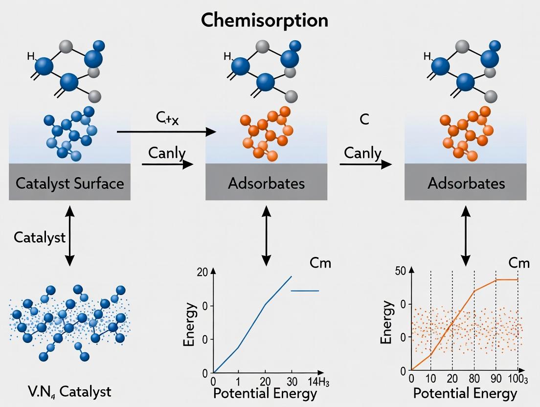
Within the broader thesis on the Fundamentals of Chemisorption for Catalyst Surface Analysis Research, understanding weak and reversible chemisorption is critical for accurately characterizing catalyst surfaces. Unlike strong, irreversible chemisorption, weak interactions are highly sensitive to experimental conditions, particularly probe molecule selection and temperature. This guide provides an in-depth technical framework for designing reliable adsorption experiments to quantify these elusive sites.
Defining Weak vs. Reversible Chemisorption
Weak Chemisorption involves adsorption energies typically between 20-50 kJ/mol, sharing characteristics with both physisorption and strong chemisorption. Reversible Chemisorption can refer to weak chemisorption or to strongly chemisorbed species that desorb intact under certain conditions (e.g., temperature-programmed desorption). The operational boundary is often defined by the experimental temperature window and the chosen probe.
Core Principles for Probe Molecule Selection
The ideal probe molecule must selectively and reversibly interact with the target surface sites without undergoing side reactions.
- Acid-Base Properties: Use basic probes (e.g., NH₃, pyridine) for acid sites; acidic probes (e.g., CO₂, SO₂) for basic sites.
- Steric Accessibility: Small molecules (CO, NO) access micropores and dense sites; larger molecules (2,6-di-tert-butylpyridine) are selective for external or strong acid sites.
- Thermal Stability: The probe must not decompose or react (e.g., disproportionate, polymerize) at the analysis temperature.
- Spectroscopic Handle: Possession of a distinct signature for IR, Raman, or NMR spectroscopy is highly advantageous.
Quantitative Guide to Probe Molecules & Temperature Ranges
The following table summarizes key probe molecules, their primary applications, and recommended temperature ranges for studying weak/reversible chemisorption.
Table 1: Probe Molecules for Weak/Reversible Chemisorption Studies
| Probe Molecule | Target Site Type | Typical Adsorption Enthalpy (kJ/mol) | Recommended Temperature Range for Reversible Binding | Key Detection Method | Notes & Cautions |
|---|---|---|---|---|---|
| Carbon Monoxide (CO) | Lewis acid sites, metal cations, metallic clusters | 30 - 80 | 77 K - 150 K (for weak sites) | IR (νCO), Microcalorimetry | Excellent for site heterogeneity. Low temps required for reversibility on many oxides. |
| Nitrogen (N₂) | Strong Lewis acid sites (e.g., Al³⁺ in zeolites) | 20 - 50 | 77 K | IR, Volumetry | Very weak, requires cryogenic temps. Highly selective for strongest sites. |
| Carbon Dioxide (CO₂) | Basic sites, alkaline earth oxides | 30 - 60 | 298 K - 373 K | IR (ν₃ asym.), TPD-MS | Can form carbonates (irreversible). Linear vs. bent coordination indicates site strength. |
| Ammonia (NH₃) | Brønsted & Lewis acid sites | 50 - 120 | 373 K - 473 K (for reversible) | TPD-MS, IR, Microcalorimetry | Often too strong; use lower temperatures (<373 K) to isolate reversible component. |
| Pyridine (C₅H₅N) | Brønsted & Lewis acid sites | ~100 | 423 K - 523 K (for reversible) | IR (fingerprint region) | Desorbs ~423K from weak Lewis sites; >523K from strong Brønsted sites. Steric hindrance adjustable. |
| Methanol (CH₃OH) | Acid-base pair sites, hydroxyl groups | 40 - 80 | 300 K - 400 K | IR, SS NMR | Can dissociate or form methoxy species. Useful for probing bifunctionality. |
| Ethene (C₂H₄) | Alkaline earth cations, weak metal sites | 40 - 70 | 200 K - 300 K | IR (π-complex), TPD | Polymerization risk on strong acid/metal sites. Good for very weak cation-π interactions. |
Experimental Protocol: Temperature-Programmed Desorption (TPD) with Reversible Probes
This protocol details a generalized method for quantifying weakly chemisorbed species via TPD.
5.1. Materials & Pre-Treatment
- Catalyst Sample: 50 - 100 mg, sieved to 250-425 µm.
- Probe Gas: High purity (≥99.95%), with appropriate drying trap (e.g., molecular sieve for CO).
- Inert Gas: Ultra-high purity He or Ar, further purified by oxygen/moisture traps.
- Reactor: Quartz U-tube or micro-reactor with frit.
- Detection: Mass Spectrometer (MS) calibrated for the probe's main m/z fragment and possible decomposition products.
5.2. Step-by-Step Workflow
- In-situ Pretreatment: Heat sample in inert flow (e.g., 20 mL/min He) to activation temperature (e.g., 773 K for 1 h) to clean the surface. Cool to desired adsorption temperature (T_ads).
- Saturation & Purge: Expose to probe gas (e.g., 5% CO/He) at T_ads for 30-60 min. Switch to inert gas at the same temperature for 60-120 min to remove all physisorbed and gas-phase species.
- Temperature Ramp: Initiate a linear temperature ramp (β = 5-20 K/min) in inert flow to the final temperature (e.g., 773 K). Continuously monitor effluent with MS.
- Data Analysis: Quantify desorption peaks by integrating the MS signal. Use a calibrated MS or downstream TCD for absolute quantification. Analyze peak shape and temperature (Tp) to estimate activation energy of desorption (Ed) using the Redhead method (for first-order kinetics).
5.3. The Scientist's Toolkit: Research Reagent Solutions
Table 2: Essential Materials for Reversible Chemisorption Experiments
| Item | Function | Critical Consideration |
|---|---|---|
| Quartz Wool & Reactor Tubes | To hold catalyst bed, inert at high temperature. | Must be pre-cleaned at temperatures exceeding use to avoid contaminant outgassing. |
| Mass Spectrometer (MS) | For sensitive, selective detection of desorbing species and decomposition fragments. | Requires careful calibration and background subtraction for quantitative work. |
| Cryogenic Thermostat | To precisely control adsorption at sub-ambient temperatures (e.g., 77 K, 100 K). | Enables use of very weak probes like N₂. Use of isopropanol/dry ice or liquid N₂ baths. |
| In-line Moisture/Oxygen Traps | To purify carrier and probe gases to ppb levels. | Prevents oxidation or hydroxylation of active surfaces during measurement. |
| Calibrated Pulse Doser | For titrating precise amounts of probe molecule in volumetric or pulse chemisorption. | Allows construction of adsorption isotherms at fixed temperature. |
| FTIR Spectroscopy Cell | For in-situ characterization of adsorbed species' identity and bonding. | Must allow precise temperature control, gas flow, and high-throughput IR windows (e.g., KBr, CaF₂). |
Logical Decision Framework for Experimental Design
The following diagram outlines the systematic decision process for selecting probe and temperature based on research goals.
Diagram Title: Decision Flow for Probe & Temperature Selection
Case Study: Differentiating Weak Lewis Acid Sites on γ-Al₂O₃
7.1. Experimental Workflow This case study visualizes the integrated workflow combining volumetric adsorption and in-situ spectroscopy.
Diagram Title: Workflow for Site-Energy Correlation Study
7.2. Interpretation: The low-temperature (100 K) CO isotherm provides the total volume of weak sites. Simultaneously, IR spectroscopy reveals distinct carbonyl stretching frequencies (e.g., 2230-2190 cm⁻¹ for Al³⁺, 2180-2150 cm⁻¹ for cationic sites). Correlating the IR intensity of specific bands with adsorbed amount allows for the construction of site-specific isotherms. Subsequent temperature increase (Step 5) monitors the reversible desorption from each spectroscopically identified site.
Mastering the study of weak and reversible chemisorption is foundational to a complete surface analysis thesis. Success hinges on the strategic pairing of a selective, thermally stable probe molecule with a precisely controlled temperature protocol. The methodologies outlined here—centered on TPD, volumetric isotherms, and in-situ spectroscopy—provide a rigorous framework to quantify and qualify these critical, yet often overlooked, catalytic sites, leading to more rational catalyst design.
Addressing Diffusion Limitations and Mass Transfer Issues in Porous Catalysts
The analysis of catalyst surfaces via chemisorption is a cornerstone of heterogeneous catalysis research, providing critical data on active site density, dispersion, and particle size. However, the fundamental premise of these measurements—that reactant molecules can freely access and chemisorb onto all active sites—is frequently invalidated by diffusion limitations within porous catalyst architectures. This whitepaper examines the interplay between mass transfer phenomena and intrinsic chemisorption kinetics, framing it as an essential correction factor for accurate surface analysis in catalyst and porous material development for chemical and pharmaceutical synthesis.
Core Mass Transfer Regimes and Their Impact
Mass transfer in porous catalysts occurs through multiple, often serial, steps. The dominance of a particular regime is determined by the pore structure and reaction conditions.
Table 1: Mass Transfer Regimes in Porous Catalysts
| Regime | Typical Pore Size (nm) | Dominant Transport Mechanism | Impact on Observed Chemisorption & Reaction Rate |
|---|---|---|---|
| Bulk Diffusion | > 1000 | Molecular & Knudsen Diffusion | Minor limitations; measured rate ≈ intrinsic rate. |
| Knudsen Diffusion | 2 - 100 | Molecule-pore wall collisions | Significant limitation; measured rate < intrinsic rate. |
| Configurational Diffusion | < 2 | Activated surface diffusion | Severe limitation; strong dependence on molecule shape. |
| Intraparticle Diffusion | N/A | Diffusion within the particle pore network | Rate controlled by pore geometry and tortuosity. |
| Interfacial Diffusion | N/A | Film diffusion across stagnant layer | Can limit access to particle exterior. |
Diagnostic Experiments and Protocols
Accurate surface analysis requires diagnostics to detect and quantify mass transfer intrusions.
Weisz-Prater Criterion for Internal Diffusion
Objective: Determine if internal diffusion limits the observed reaction rate during a catalytic test preceding chemisorption analysis. Protocol:
- Conduct a steady-state reaction in a differential reactor under known conditions.
- Measure the observed reaction rate, ( r_{obs} ) (mol·g⁻¹·s⁻¹).
- Determine the catalyst particle radius, ( R ) (cm), and estimate the effective diffusivity, ( D_{eff} ) (cm²·s⁻¹).
- Calculate the Weisz-Prater modulus, ( C{WP} = \frac{r{obs} \rho{cat} R^2}{D{eff} Cs} ), where ( \rho{cat} ) is pellet density (g·cm⁻³) and ( C_s ) is surface concentration (mol·cm⁻³).
- Interpretation: If ( C{WP} \ll 1 ), no internal diffusion limitations. If ( C{WP} \gg 1 ), severe limitations exist, and chemisorption measurements may undercount active sites.
Koros-Nowak (Madon-Boudart) Test for External Diffusion
Objective: Verify the absence of external (film) mass transfer limitations. Protocol:
- Perform catalytic rate measurements at constant temperature and reactant partial pressure while varying the catalyst mass (( m )) and total gas flow rate (( F )) such that the contact time (( m/F )) remains constant.
- Plot the observed rate versus total catalyst mass.
- Interpretation: A constant observed rate independent of catalyst mass indicates the absence of external diffusion limitations. A decreasing rate with increasing mass suggests limitations.
Pulse Chemisorption with Varied Particle Size
Objective: Directly probe intraparticle diffusion effects on chemisorption measurement. Protocol:
- Sieve the catalyst sample into several distinct particle size fractions (e.g., 150-250 µm, 45-75 µm).
- For each fraction, perform dynamic pulse chemisorption (e.g., H₂ or CO) using a Micromeritics ASAP 2920 or equivalent.
- Measure the total chemisorbed gas per gram of catalyst for each fraction.
- Interpretation: Constant uptake across all sizes indicates diffusion-free measurement. A decrease in uptake with increasing particle size signals diffusion limitations, rendering the chemisorption data unreliable for dispersion calculation.
Diagram: Protocol for Particle-Size Dependent Chemisorption
Advanced Techniques to Overcome Limitations
Designing Hierarchical Pore Networks
Strategy: Introduce macro- or mesopores as transport highways to mitigate diffusion barriers in microporous active components. Synthesis Protocol (Soft-Templating for Meso/Macroporous Zeolites):
- Prepare a precursor gel containing zeolite structure-directing agents (e.g., tetrapropylammonium hydroxide for ZSM-5) and a silica source (e.g., tetraethyl orthosilicate).
- Add a soft template (e.g., amphiphilic polymer like Pluronic P123) and stir vigorously for 24h.
- Transfer the gel to an autoclave for hydrothermal crystallization at 150-180°C for 24-72h.
- Cool, filter, wash, and dry the solid.
- Calcine in air at 550°C for 6h to remove organic templates.
Table 2: Impact of Hierarchical Porosity on Catalytic Performance
| Catalyst Type | Total Surface Area (m²/g) | Mesopore Volume (cm³/g) | Apparent Rate Constant (k_obs) for Test Reaction | Thiele Modulus (Estimated) |
|---|---|---|---|---|
| Conventional Zeolite Y | 780 | 0.05 | 1.0 (Baseline) | 4.2 |
| Hierarchical Zeolite Y | 650 | 0.35 | 3.8 | 1.1 |
Thin-Film Catalysts and Monoliths
Strategy: Coat active phases as thin films (<50 µm) onto monolithic supports (cordierite, alumina) to drastically shorten intraparticle diffusion paths. Washcoating Protocol:
- Prepare a stable slurry of catalyst powder (d90 < 10 µm) in deionized water. Adjust pH with nitric acid or ammonia to achieve a zeta potential > |30| mV for stability.
- Add a binder (e.g., colloidal alumina, 5 wt% of solid) and stir.
- Immerse the clean monolith substrate, withdraw at a controlled rate (e.g., 2 cm/min), and blow excess slurry from channels with air.
- Dry at 110°C for 2h and calcine at 450°C for 4h to adhere the washcoat.
- Repeat to achieve desired loading (typically 10-20 wt%).
The Scientist's Toolkit: Research Reagent Solutions
Table 3: Essential Materials for Diffusion and Chemisorption Studies
| Item | Function & Rationale |
|---|---|
| Micromeritics ASAP 3Flex | Physisorption analyzer for full BET surface area and pore size distribution (PSD) analysis via N₂ at 77K. Critical for characterizing diffusion pathways. |
| Micromeritics AutoChem II | Chemisorption analyzer for pulse or TPD/TPR studies using H₂, CO, O₂, etc. Determines active metal surface area and dispersion. |
| Pluronic P123 Triblock Copolymer | Soft template for synthesizing ordered mesoporous silica (SBA-15) or hierarchical zeolites via evaporation-induced self-assembly. |
| Cetyltrimethylammonium Bromide (CTAB) | Surfactant template for synthesizing MCM-41 mesoporous silica with tunable pore diameter (2-5 nm). |
| Tetraethyl Orthosilicate (TEOS) | Common silica precursor for sol-gel synthesis of controlled-porosity materials. |
| Trimethylaluminum (TMA) | Aluminum precursor for Atomic Layer Deposition (ALD) to create uniform active sites or diffusion barriers in pores. |
| Sieving Kit (ASTM Mesh Series) | For precise particle size fractionation (e.g., 45, 75, 150 µm) to perform diffusion diagnostics. |
| Cordierite Monolith (400 cpsi) | Standard ceramic support for washcoating to create diffusion-shortened catalytic reactors. |
| Colloidal Alumina Binder (20% wt) | Stabilizes catalyst washcoats on monoliths, preventing peel-off during reaction while adding minimal diffusion resistance. |
Diagram: Diagnostic & Mitigation Workflow for Mass Transfer Issues
Overcoming Problems with Non-Stoichiometric or Complex Adsorption Stoichiometries
Within the broader thesis on the Fundamentals of Chemisorption for Catalyst Surface Analysis Research, the precise determination of adsorption stoichiometry is paramount. Non-stoichiometric or complex adsorption stoichiometries—where the adsorbate-to-surface-site ratio is not a simple integer or varies with coverage, pressure, or temperature—present significant challenges. These complexities obscure critical metrics like active site density, turnover frequencies, and structure-activity relationships. This guide provides a technical framework for identifying, characterizing, and overcoming these challenges using modern surface science and spectroscopic methodologies.
Core Challenges and Diagnostic Signs
Non-stoichiometric behavior arises from several phenomena:
- Adsorbate-induced Surface Reconstruction: Adsorption triggers surface atom rearrangement, changing the number and type of available sites.
- Multidentate or Dissociative Adsorption: A single molecule binds to multiple sites or dissociates into fragments that adsorb independently.
- Lateral Interactions: Repulsive or attractive interactions between adsorbed species alter the effective saturation coverage.
- Heterogeneous Surfaces: The presence of multiple distinct surface facets, defects, or impurities with different binding energies and capacities.
- Precursor-Mediated Adsorption: Physisorbed states lead to kinetic complexities that mask true chemisorption equilibria.
Diagnostic data indicating complex stoichiometry include:
- Non-linear or multi-step Langmuir isotherms.
- Coverage-dependent enthalpies of adsorption measured by calorimetry.
- Multiple desorption peaks in temperature-programmed desorption (TPD) not attributable to simple desorption kinetics.
- Shifts in vibrational frequencies (e.g., in FTIR) as a function of coverage.
Table 1: Common Adsorbates Exhibiting Complex Stoichiometry on Metal Surfaces
| Adsorbate | Typical Surface(s) | Apparent Stoichiometry Range (Molecule:Site) | Primary Complexity Cause |
|---|---|---|---|
| Carbon Monoxide (CO) | Pt(111), Rh(111) | 0.2 - 0.8 | Bridging vs. linear bonding, island formation. |
| Nitric Oxide (NO) | Pt(100), Pd(111) | 0.3 - 1.0 | Dissociation (N,O) vs. molecular adsorption. |
| Ammonia (NH₃) | Cu(110), SiO₂-supported Ni | 0.1 - 1.0 | Hydrogen-bonding networks, desorption with decomposition. |
| Ethylene (C₂H₄) | Ni(111), Pt(111) | 0.15 - 0.5 | Dehydrogenation to ethylidyne (CCH₃) or dissociation. |
| Hydrogen (H₂) | Most transition metals | 0.5 - 2.0 | Dissociative adsorption, subsurface penetration. |
Table 2: Comparison of Techniques for Resolving Complex Stoichiometries
| Technique | Key Measurable | Stoichiometry Insight | Practical Limitations |
|---|---|---|---|
| Static Chemisorption (Volumetric) | Uptake at defined P, T | Site density if stoichiometry is known. | Assumes simple 1:1 model; fails for complexes. |
| TPD/MS | Desorption energy, peak profile | Identifies distinct adsorbed states & relative populations. | Requires careful calibration; can be kinetic-limited. |
| Microcalorimetry | ΔH_ads vs. Coverage | Direct measure of site heterogeneity & interaction strength. | Experimentally demanding; low surface area samples. |
| In-situ FTIR/DRIFTS | Vibrational mode shifts | Identifies binding configurations (e.g., linear vs. bridged CO). | Quantification challenging; requires extinction coeff. |
| Synchrotron XPS/NAP-XPS | Core-level binding energy shifts | Oxidation state, adsorption-induced surface shifts. | Requires ultra-high vacuum or specialized cells. |
Experimental Protocols
Protocol 1: Combined TPD andIn-SituFTIR for CO Stoichiometry Determination
Objective: To deconvolute the contributions of linear (1:1) and bridged (1:2) CO adsorption on a supported metal catalyst.
Materials: See "The Scientist's Toolkit" below.
Method:
- Catalyst Pretreatment: Reduce 100 mg of catalyst (e.g., 2% Pt/Al₂O₃) in a 30 ml/min H₂ flow at 400°C for 2 hours. Purge with inert gas (He) at 400°C for 1 hour, then cool to 30°C under inert flow.
- Dosing: Expose the catalyst to a calibrated 5% CO/He mixture in pulses (via a loop injector) until saturation is reached, monitoring effluent with an online TCD.
- In-Situ FTIR Measurement: Transfer a separate, identically pretreated waferized sample to an in-situ DRIFTS cell. After identical reduction and purge, collect a background spectrum at 30°C under He. Introduce 1% CO/He and collect spectra sequentially (4 cm⁻¹ resolution) as a function of time to saturation.
- TPD Sequence: Following dosing in the TPD system, flush the sample with He at 30°C for 30 minutes to remove physisorbed CO. Initiate a linear temperature ramp (e.g., 10°C/min) to 600°C under He flow, monitoring desorbing CO (m/z=28) with a calibrated mass spectrometer.
- Data Integration & Analysis:
- Integrate the total TPD peak area and convert to moles using a calibration curve from known CO pulses.
- Deconvolute the IR spectra in the C-O stretching region (1800-2100 cm⁻¹) using peak fitting for linear (~2050-2080 cm⁻¹) and bridged (~1800-1900 cm⁻¹) species.
- Assuming the extinction coefficient for linear CO is known or can be approximated from literature, estimate its relative coverage. Use the TPD-derived total moles and the IR-derived fraction to calculate the weighted average stoichiometry.
Protocol 2: Adsorption Microcalorimetry for Energetic Heterogeneity Mapping
Objective: To measure the differential heat of adsorption as a function of coverage to quantify site heterogeneity and lateral interactions.
Method:
- System Preparation: Calibrate the microcalorimeter (e.g., a Calvet-type) using electrical pulses and standard gas injections (e.g., Kr adsorption on non-porous alumina).
- Sample Activation: Place 50-100 mg of high-surface-area catalyst in the sample cell. Activate in vacuo (P < 10⁻⁶ mbar) at the required temperature (e.g., 400°C for metals) for 2 hours.
- Incremental Dosing: Isothermally control the sample at the adsorption temperature (e.g., 30°C). Introduce small, sequential doses of the probe gas (e.g., NH₃, CO) from a calibrated volume. After each dose:
- Record the thermal profile (heat flow vs. time) until baseline returns.
- Record the pressure drop in the manifold to calculate the amount adsorbed.
- Calculation: Integrate the area under each heat flow peak to obtain the integral heat (Qint) for that dose. The differential heat (Qdiff) is Qint divided by the amount adsorbed in that dose. Plot Qdiff versus total adsorbate coverage.
- Interpretation: A constant Qdiff indicates homogeneous sites. A steadily declining Qdiff indicates either intrinsic heterogeneity or repulsive lateral interactions. A sharp initial drop followed by a plateau suggests distinct sets of strong then weak sites.
The Scientist's Toolkit
Table 3: Key Research Reagent Solutions & Materials
| Item | Function / Rationale |
|---|---|
| High-Purity, Well-Defined Catalyst (e.g., Single Crystal or Synthesized Nanoparticles) | Minimizes ambiguity from unknown impurities and ill-defined facets. Enables correlation of structure to stoichiometry. |
| Ultra-High Purity Gases with In-line Purifiers/Mass Filters | Eliminates contaminants (e.g., Fe(CO)₅ in CO, O₂ in H₂) that can poison surfaces or participate in side reactions. |
| Calibrated, Leak-Tight Volumetric/Pulse Chemisorption System | Provides the fundamental quantitative uptake measurement. Accuracy is foundational for all further analysis. |
| Mass Spectrometer (QMS) with Fast Response, Calibrated for Quantification | Essential for TPD to identify desorbing species and quantify amounts via fragmentation pattern analysis. |
| In-Situ or Operando Spectroscopy Cell (DRIFTS, XAS, XPS) | Allows observation of adsorbate bonding configuration and surface state under relevant conditions. |
| Reference Adsorbate with Known, Simple Stoichiometry (e.g., H₂ on Pt, CO on Pd at low P) | Provides a calibration point for active site counting when the stoichiometry is well-established (e.g., H:Pt = 1:1). |
| Density Functional Theory (DFT) Computational Resources | Models adsorption configurations, energies, and vibrational spectra to assign experimental observations to atomic-scale structures. |
Visualizations
Diagram Title: Decision Workflow for Complex Stoichiometry Analysis
Diagram Title: Complex Adsorption Pathways for CO and NH₃
Best Practices for Sample Preparation, Reduction, and Pre-Treatment to Ensure Clean Surfaces.
In the rigorous study of chemisorption for catalyst surface analysis, the quality of data is intrinsically linked to the condition of the catalyst surface. A pristine, well-defined surface, free of contaminants and with a known oxidation state, is the fundamental prerequisite for obtaining meaningful adsorption isotherms, accurate active site counts (dispersion, metal surface area), and reliable mechanistic insights. This guide details the core practices to achieve such surfaces, framed within the thesis that meticulous pre-treatment is not a preliminary step but the cornerstone of fundamental chemisorption research.
Foundational Principles of Surface Cleaning
The objective of sample pre-treatment is to remove physisorbed species (e.g., water, atmospheric hydrocarbons, carbonates) and chemisorbed contaminants (e.g., coke, sulfur, chlorine) while reducing the active phase (typically metals) to a zero-valent or desired oxidation state without inducing sintering or structural alteration. The process is governed by the interplay of temperature, gas environment, and time.
Quantitative Pre-Treatment Parameters for Common Catalysts
The table below summarizes optimized conditions for common catalyst systems, derived from recent literature and standard protocols. These conditions are starting points and may require optimization for specific materials.
Table 1: Standard Reduction/Pre-Treatment Conditions for Catalyst Systems
| Catalyst System | Typical Pre-Treatment Goal | Temperature Range (°C) | Gas Environment & Flow | Duration (hours) | Critical Considerations |
|---|---|---|---|---|---|
| Supported Noble Metals (Pt, Pd, Rh/Al₂O₃, SiO₂) | Reduction to metallic state | 300 - 450 | 5% H₂/Ar or 100% H₂, 30-50 mL/min | 2 - 4 | Lower T for high dispersion; avoid H₂ spillover on reducible supports. |
| Supported Base Metals (Ni, Co, Cu/Al₂O₃, SiO₂) | Reduction to metallic state | 350 - 500 | 5-10% H₂/Ar, 30-50 mL/min | 3 - 6 | Higher T often needed; monitor for reduction completeness via TPR. |
| Reducible Oxide Catalysts (Cu/ZnO, CeO₂-based) | Surface reduction/activation | 200 - 300 | 5% H₂/Ar or CO, 20-30 mL/min | 1 - 2 | Goal is often a partially reduced surface; bulk reduction can be detrimental. |
| Sulfided Catalysts (Co-Mo/Al₂O₃) | Pre-sulfidation | 350 - 400 | 10% H₂S/H₂ or CS₂/H₂, 30 mL/min | 2 - 4 | Requires specialized equipment for safe handling of sulfiding agents. |
| Cleaning after Reaction (Coke Removal) | Oxidative regeneration | 450 - 550 (max) | 2-5% O₂/He or air, 30 mL/min | 2 - 6 | Slow ramps to avoid runaway exotherms; may alter metal dispersion. |
Detailed Experimental Protocols
Protocol for StandardIn-SituReduction of a Supported Metal Catalyst
This protocol precedes volumetric or dynamic chemisorption analysis (e.g., H₂ or CO pulse chemisorption).
Objective: To reduce surface metal oxides to the metallic state and remove surface contaminants. Materials: Quartz/U-shaped sample cell, mass flow controllers, temperature-programmed furnace, thermal conductivity detector (TCD) optional. Reagent Solutions: Ultra-high purity (UHP) H₂ (5% in Ar), UHP Argon. Safety: Ensure proper ventilation for H₂ use; perform leak checks.
Procedure:
- Loading: Precisely weigh (typically 50-200 mg) catalyst into the sample cell. Plug with quartz wool.
- Initial Purge: Mount the cell in the manifold. At room temperature, purge with inert gas (Ar, 30 mL/min) for 30 minutes to displace air.
- Temperature Ramp: Under continuous inert flow, ramp temperature to 150°C at 10°C/min. Hold for 60 minutes to remove physisorbed water and volatile organics.
- Switch to Reducing Gas: Switch gas flow to 5% H₂/Ar (30 mL/min). Allow gas to stabilize for 10 minutes at 150°C.
- Reduction Ramp: Increase temperature to the target reduction temperature (e.g., 400°C for Pt/Al₂O₃) at 5-10°C/min.
- Reduction Hold: Maintain at the target temperature for 2-4 hours under reducing flow.
- Cooling & Purging: Cool the sample to the analysis temperature (often 35-50°C for H₂ chemisorption) under the reducing gas flow. Once at temperature, switch to inert gas (Ar) and purge for 30-60 minutes to remove any weakly adsorbed hydrogen. Critical: The sample must never be exposed to air after this point.
- Proceed to Analysis: The catalyst is now ready for chemisorption measurements.
Protocol for Oxidative Cleaning (Decoking)
Used to regenerate catalysts fouled by carbonaceous deposits.
Procedure:
- After reaction, cool the sample to <100°C under inert gas.
- Switch to a mild oxidizing mixture (2% O₂ in He, 30 mL/min).
- Ramp temperature slowly (2-3°C/min) to 450°C. Monitor TCD signal for CO₂ production peaks.
- Hold at 450°C until the CO₂ signal returns to baseline (typically 2-6 hours).
- Cool to reduction temperature in inert gas, then follow the standard reduction protocol (Section 4.1) to re-reduce the metal phase.
Visualization of Workflows
Diagram 1: Universal Catalyst Pre-Treatment Workflow (76 chars)
Diagram 2: Mechanism of Surface Reduction via H₂ (63 chars)
The Scientist's Toolkit: Essential Research Reagent Solutions
Table 2: Key Materials for Surface Preparation and Analysis
| Reagent/Material | Typical Purity | Primary Function in Pre-Treatment |
|---|---|---|
| 5% H₂ in Ar (Balance Gas) | UHP (>99.999%) | Standard reducing mixture for safe, controlled metal oxide reduction. |
| Ultra-High Purity Argon/Helium | UHP (>99.999%) | Inert purge gas for drying, cooling, and carrier gas in analysis. |
| 2-10% O₂ in He | UHP (>99.999%) | Mild oxidizing mixture for controlled removal of carbonaceous deposits. |
| Carbon Monoxide (CO) | UHP (>99.97%) | Reducing agent for specific oxides; probe molecule for chemisorption. |
| Quartz Wool & Sample Tubes | High-purity, fused quartz | Inert sample containment; must be pre-cleaned at high temperature. |
| Molecular Sieves (3Å, 5Å) | Activated | Used in gas purification trains to remove trace water from gas lines. |
| Oxytraps & Hydrocarbontraps | Lab-specific | In-line filters to remove O₂ and hydrocarbon impurities from gas streams. |
Validating Chemisorption Data: Cross-Technique Correlation and Advanced Surface Analysis
Within the thesis on Fundamentals of Chemisorption for Catalyst Surface Analysis Research, a central challenge is validating the volumetric, ensemble-averaged surface area and active site density derived from chemisorption with direct, spatially-resolved observations of catalyst nanoparticles. Transmission Electron Microscopy (TEM) and Scanning Transmission Electron Microscopy (STEM) provide the requisite nanoscale imaging to measure particle size distributions (PSDs). Correlating these two disparate data sets is critical for determining structure-activity relationships, dispersion calculations, and validating the assumptions inherent in chemisorption models (e.g., uniform particle morphology and stoichiometric adsorbate-to-metal ratios).
Fundamentals: Chemisorption and Microscopy
Chemisorption Analysis (Volumetric/Manometric Method): This technique measures the quantity of a selective gas (e.g., H₂, CO, O₂) that chemically bonds to the active metal surface at equilibrium conditions. The total uptake is used to calculate:
- Metal Dispersion (D): D = (Number of surface metal atoms / Total number of metal atoms) × 100%
- Average Particle Size (d_Chem): Assuming a specific geometric model (typically spherical cuboctahedra), d_Chem (nm) = k / D, where k is a shape-dependent constant (~0.9-1.1 for many metals with H₂ chemisorption).
Electron Microscopy (TEM/STEM): Provides direct images of particles. Statistical analysis of hundreds of particles yields a number-based Particle Size Distribution (PSD), from which key metrics are derived:
- Number-Average Diameter (dₙ): Σ(nᵢdᵢ) / Σnᵢ
- Surface-Average Diameter (dₛ): Σ(nᵢdᵢ³) / Σ(nᵢdᵢ²) — This is directly comparable to d_Chem, as chemisorption measures surface-area-weighted properties.
Quantitative Correlation: Data Comparison Table
The core of the correlation lies in comparing the area-weighted average size from microscopy with the chemisorption-derived size.
Table 1: Comparative Metrics from Chemisorption and Electron Microscopy
| Metric | Chemisorption (Volumetric) | TEM/STEM (Image Analysis) | Correlation Principle |
|---|---|---|---|
| Primary Output | Total gas uptake (μmol/g) | Lattice-resolved or Z-contrast images | – |
| Key Assumption | Adsorbate:metal stoichiometry, uniform site reactivity, particle geometry | 2D projection represents 3D object, thresholding accuracy | Both methods assume representative sampling of the bulk catalyst. |
| Calculated Size | d_Chem (surface-mean diameter) | dₛ (surface-area-weighted mean diameter) | d_Chem ≈ dₛ for perfect correlation. |
| Size Distribution | Indirect, inferred from model (mono- vs. poly-disperse) | Direct PSD (histogram from n>200 particles) | PSD explains deviations; a long tail of large particles increases dₛ over dₙ. |
| Information Depth | Bulk powder average (mg) | Local, ultra-thin region (pg) | Multiple microscopy fields required for statistical significance. |
Table 2: Common Discrepancies and Their Origins
| Observation | Probable Cause | Implications for Thesis Research |
|---|---|---|
| d_Chem > dₛ | Inaccessible/passivated surface atoms, strong metal-support interactions blocking chemisorption. | Overestimation of particle size from chemisorption; true active surface area is smaller. |
| d_Chem < dₛ | Chemisorption on support or metal cations, stoichiometry error (e.g., H/M >1), non-spherical particles. | Overestimation of metal dispersion; particle counting may miss very small clusters (<1 nm). |
| Poor Correlation | Bimodal PSD, non-uniform adsorbate bonding, poor microscopy sample prep (aggregation). | Highlights limitation of using only average metrics; full PSD analysis is essential. |
Experimental Protocols for Correlation
Protocol 1: Static Volumetric Chemisorption for Dispersion
- Sample Preparation: ~50-100 mg of reduced/passivated catalyst is loaded into a quartz cell. The sample is degassed under vacuum (<10⁻⁵ mbar) at 150°C for 1 hour to remove physisorbed species.
- Reduction In Situ: The sample is heated under flowing H₂ (typically 5% in Ar) at a specified temperature (e.g., 350°C for 2 hours) to reduce the metal phase, followed by evacuation at the reduction temperature for 1 hour.
- Adsorption Isotherm: The sample cell is immersed in a liquid N₂ bath (77 K). Known doses of the probe gas (e.g., H₂) are introduced sequentially. The equilibrium pressure after each dose is recorded.
- Data Analysis: The total uptake is determined from the linear portion of the isotherm via extrapolation to zero pressure. Dispersion (D) and d_Chem are calculated using assumed stoichiometry (e.g., H:Pt=1:1) and particle geometry.
Protocol 2: TEM/STEM Sample Prep and Image Analysis for PSD
- Dry Dispersion: Catalyst powder is lightly ground and dispersed in ethanol via sonication for 30-60 seconds. A drop is deposited on a lacey carbon/Cu grid and dried.
- Microscopy Acquisition: Imaging is performed at appropriate magnification (e.g., 400k-800kx) to resolve particle boundaries. For STEM-HAADF, contrast is approximately proportional to Z².
- Image Analysis Workflow:
- Thresholding: Adjust contrast to separate particles from background.
- Particle Identification: Use software (e.g., ImageJ, DigitalMicrograph) to detect individual particles. Manual verification is critical.
- Measurement: Record Feret's diameter or equivalent circular diameter for each particle (n > 200).
- Statistical Calculation: Generate histogram and calculate dₙ, dₛ, and standard deviation.
Visualization of the Correlation Workflow
Title: Chemisorption-Microscopy Correlation Workflow
Title: Key Metrics for Size Correlation
The Scientist's Toolkit: Essential Research Reagents & Materials
Table 3: Key Reagents and Materials for Correlation Studies
| Item | Function & Specification | Critical Note |
|---|---|---|
| Probe Gases | H₂ (Ultra-high purity, 99.999%): For noble metal (Pt, Pd) dispersion. CO (99.99%): For metals like Ru, Fe. O₂ (99.99%): For titration or base metal analysis. | Must be further purified with traps to remove trace O₂/H₂O. Stoichiometry assumption is critical. |
| Catalyst Sample Cells | Quartz or glass U-tube/triple-port cells with high-vacuum stopcocks. | Must withstand in situ reduction temperatures and high vacuum. |
| TEM Grids | Lacey carbon film on 300-mesh Cu or Au grids. | Au grids avoid Cu signal interference in EDS. Lacey carbon provides thin support. |
| Dispersion Solvent | Anhydrous Ethanol or Isopropanol (HPLC grade). | Low surface tension, volatile, and does not react with the catalyst. |
| Reference Material | Certified nanoparticle size standard (e.g., Au nanoparticles on carbon). | For daily verification of TEM/STEM magnification calibration. |
| Image Analysis Software | Fiji/ImageJ (open-source) or DigitalMicrograph (commercial). | Essential for batch processing and statistical analysis of PSD. |
| High-Vacuum System | Adsorption analyzer or custom-built volumetric/manometric setup with pressure transducers (0-1000 Torr). | Base pressure <10⁻⁵ mbar is required for accurate uptake measurements. |
This whitepaper, framed within a broader thesis on the Fundamentals of Chemisorption for Catalyst Surface Analysis Research, addresses a critical challenge in heterogeneous catalysis: quantifying active surface sites from elemental composition data. While X-ray Photoelectron Spectroscopy (XPS) and Auger Electron Spectroscopy (AES) provide superb quantitative analysis of surface composition (typically the top 1-10 nm), they do not directly measure the number of catalytically active sites. This guide details a methodology to correlate XPS/AES-derived composition with active site densities obtained from chemisorption techniques, enabling a more profound understanding of structure-activity relationships.
Core Principles: Bridging Composition to Site Density
The active site count is typically measured via selective chemisorption (e.g., H₂, CO, N₂O titration) coupled with volumetric or flow apparatus. The fundamental link to XPS/AES data is established through the following relationship:
Active Site Density (Sites/cm²) = [Surface Atom Density (Atoms/cm²)] × [Surface Fraction (from XPS)] × [Dispersion Factor]
Where:
- Surface Atom Density is derived from the known crystal structure and morphology (e.g., ~1.3×10¹⁵ Pt atoms/cm² for Pt(111)).
- Surface Fraction is the quantitative concentration of the catalytic element from XPS (e.g., Pt 4f peak area, corrected by sensitivity factors).
- Dispersion Factor is the fraction of surface atoms that are active, often initially assumed from chemisorption and refined by correlation.
Experimental Protocols
Protocol A: Integrated XPS & Chemisorption Measurement on a Single Sample
Objective: To obtain surface composition and active site count from the exact same sample spot, minimizing heterogeneity errors.
- Sample Preparation: Deposit catalyst powder onto a dedicated, ultra-high vacuum (UHV) compatible sample stub using conductive adhesive. Alternatively, use a foil or thin film model catalyst.
- Initial XPS/AES Analysis:
- Load sample into UHV system (base pressure < 5×10⁻⁹ mbar).
- Acquire survey spectrum (0-1200 eV) to identify all elements.
- Acquire high-resolution spectra for catalytic element(s) (e.g., Pt 4f, Co 2p, Ni 2p), support elements (e.g., Al 2p, Si 2p, O 1s), and any contaminants (C 1s).
- Optional: Perform AES mapping on a selected region to confirm homogeneity.
- Use Shirley or Tougaard background subtraction. Quantify using relative sensitivity factors (RSFs) provided by the instrument manufacturer or from standard databases.
- In-situ Chemisorption (Requires UHV-Integrated System):
- Isolate analysis chamber. Introduce ultra-pure probe gas (e.g., H₂, CO) at a controlled pressure (typically 1×10⁻⁶ to 1×10⁻³ mbar).
- Monitor the surface using XPS or AES in real-time. The chemisorption event is indicated by a shift in the binding energy of the adsorbate (e.g., O 1s for CO) or the substrate metal (due to final state effects).
- The saturation coverage is determined when the adsorbate signal stabilizes. The absolute number of adsorbed molecules is calibrated against a standard.
- Post-Chemisorption XPS/AES:
- Pump out probe gas.
- Repeat high-resolution XPS on the same region. This confirms the adsorbate identity and checks for any reduction/oxidation of the surface during exposure.
- Data Correlation: Active site density from step 3 is directly correlated with the pre-adsorption surface composition from step 2.
Protocol B: Ex-situ Correlation Using Multiple Samples from a Uniform Batch
Objective: To establish a statistical correlation between surface composition and site density for a catalyst series.
- Batch Synthesis: Synthesize a well-defined series of catalysts (e.g., varying metal loading, promoter concentration) with high uniformity.
- Divided Sample Analysis:
- Split each catalyst batch into two (or more) aliquots.
- Aliquot 1: Analyze via standard volumetric/temperature-programmed chemisorption (TPC) to determine total active site count per gram of catalyst.
- Aliquot 2: Prepare for XPS/AES analysis as in Protocol A, Step 1. Analyze multiple spots/particles to obtain a statistically representative surface composition.
- Normalization: Normalize the chemisorption site count (per gram) to the catalyst's specific surface area (from BET measurements) to calculate site density (sites/m²). Correlate this value with the surface atomic percent of the active component from XPS.
Data Presentation & Correlation
Table 1: Exemplar Data Correlation for a Pt/γ-Al₂O₃ Catalyst Series
| Catalyst ID | XPS Pt 4f Atomic % (Surface) | XPS Al 2p / Pt 4f Ratio | Chemisorption (H₂ Uptake) (μmol/g) | BET SSA (m²/g) | Calculated Site Density (Sites/m²) ×10¹⁷ | Pt Dispersion (%) (from H₂) |
|---|---|---|---|---|---|---|
| PtAl-1 | 0.95 | 42.1 | 112.4 | 154 | 4.39 | 58.2 |
| PtAl-2 | 1.82 | 21.8 | 198.7 | 152 | 7.87 | 62.1 |
| PtAl-3 | 3.15 | 12.5 | 315.2 | 148 | 12.8 | 59.5 |
| PtAl-4 | 4.20 | 9.3 | 381.5 | 145 | 15.8 | 52.9 |
Table 2: The Scientist's Toolkit: Key Research Reagent Solutions & Materials
| Item | Function/Description | Critical Specification |
|---|---|---|
| Conductive Adhesive Tape (Carbon-based) | To mount powder catalysts for XPS/AES without introducing interfering signals. | High-purity, UHV-compatible, low outgassing. |
| Certified Reference Materials (e.g., Au, Cu, Ag foils) | For binding energy scale calibration and spectrometer function verification. | Traceably calibrated, clean, polished. |
| Ultra-Pure Probe Gases (H₂, CO, O₂) | For in-situ or sequential chemisorption experiments. | 99.999% purity, with in-line purifiers/mass spec. |
| Ion Sputtering Source (Ar⁺) | For gentle surface cleaning or depth profiling to assess homogeneity. | Precision current control (µA range), rasterable. |
| Relative Sensitivity Factor (RSF) Library | Database for converting XPS peak areas to atomic concentrations. | Validated for your specific spectrometer and settings. |
| Model Catalyst Wafer | Well-defined single crystal or thin film used as a calibration standard. | Known surface orientation and atom density. |
Visualization of Methodologies
Protocol A: In-situ Correlation Workflow
Protocol B: Ex-situ Statistical Correlation
Within the fundamental research on chemisorption for catalyst surface analysis, characterizing the catalyst's physical and chemical surface is paramount. While chemisorption probes active sites, physisorption, particularly Brunauer-Emmett-Teller (BET) analysis, quantifies the total specific surface area (SSA) and pore architecture. This whitepaper details the synergistic integration of these techniques, arguing that a comprehensive understanding of catalytic performance requires correlating the total SSA (from BET) with the active surface area (from chemisorption). We provide current experimental protocols, data interpretation frameworks, and visual workflows to guide researchers in obtaining a complete picture of heterogeneous catalysts and advanced materials.
In catalysis research, the total surface area, measured via physisorption of inert gases like N₂ at 77 K, represents the landscape available for reaction. However, only a fraction of this landscape—the active surface area—comprises specific sites (e.g., metal atoms, acid centers) where chemisorption and catalysis occur. Relying solely on BET area can be misleading; a high SSA does not guarantee high activity if the active site density is low. Conversely, a material with moderate SSA but exceptionally high active site density can outperform. Thus, the synergy lies in combining these metrics to calculate active site dispersion, turnover frequencies (TOF), and structure-activity relationships.
Foundational Principles
BET Physisorption Theory (Total SSA)
The BET theory models multilayer adsorption on a non-porous or macro/mesoporous solid. The linearized BET equation is applied within a relative pressure (P/P₀) range typically of 0.05–0.30: [ \frac{P}{n(P0 - P)} = \frac{1}{nm C} + \frac{C - 1}{nm C} \left( \frac{P}{P0} \right) ] Where:
- ( n ) = amount of gas adsorbed
- ( n_m ) = monolayer capacity
- ( C ) = BET constant related to adsorption energy.
The total specific surface area ( S{BET} ) is calculated from ( nm ).
Chemisorption for Active Surface Area
Chemisorption uses reactive probes (H₂, CO, O₂, NH₃) that form chemical bonds with specific surface sites. By assuming a stoichiometry (e.g., H:Metalsurface = 1:1 for H₂ chemisorption on Pt), the volume of chemisorbed gas is used to calculate:
- Active Metal Surface Area (AMSA): The surface area occupied by exposed metal atoms.
- Metal Dispersion (D): Percentage of total metal atoms exposed on the surface.
- Active Site Density: Number of active sites per unit mass or unit total area.
Experimental Protocols
Integrated Workflow for Combined Analysis
A robust characterization protocol involves sequential analysis on the same sample.
Protocol: Sequential Physisorption and Chemisorption
- Sample Preparation (~100 mg): Weigh sample in a quartz cell. Apply in-situ pretreatment.
- Pretreatment: Heat under vacuum or inert gas flow (e.g., He) to 150–300°C (for support cleaning) or higher (e.g., 400°C for metal oxide reduction in 5% H₂/Ar) for 1–2 hours, followed by evacuation.
- BET Physisorption (N₂ at 77 K):
- Cool sample to cryogenic temperature (77 K using liquid N₂ bath).
- Perform adsorption isotherm measurement across P/P₀ = 0.01–0.99.
- Apply BET equation to the 0.05–0.30 range to calculate ( S_{BET} ) and total pore volume.
- Use BJH or NLDFT methods on the desorption branch for pore size distribution.
- Degas: Re-evacuate sample at mild temperature (e.g., 150°C) to remove physisorbed N₂.
- Chemisorption (e.g., H₂ or CO Pulse Chemisorption at 35°C):
- Cool/equilibrate to analysis temperature (often 35°C for H₂).
- Expose sample to repeated small pulses of probe gas from a calibrated loop in a carrier stream (e.g., He).
- Measure unadsorbed gas with a thermal conductivity detector (TCD) until saturation.
- Calculate chemisorbed gas volume from cumulative uptake.
- Data Correlation: Calculate dispersion, active area, and site density relative to ( S_{BET} ).
Static Volumetric Method Details
For high-precision active area measurement, the static volumetric method is preferred.
- After pretreatment, the sample cell is isolated in a known volume.
- Small, precise doses of probe gas are introduced.
- The equilibrium pressure after each dose is recorded.
- The adsorbed quantity is calculated using the real gas law (e.g., Peng-Robinson) from pressure drop.
- An adsorption isotherm is constructed. The monolayer capacity is determined by extrapolation methods (e.g., Langmuir fit for uniform sites, or chemical titration for acidic sites using NH₃-TPD).
Data Presentation & Interpretation
Table 1: Comparative Data from a Model Pt/Al₂O₃ Catalyst
| Parameter | Symbol | Value | Method/Probe | Derived Metric |
|---|---|---|---|---|
| Total SSA | ( S_{BET} ) | 180 m²/g | N₂ Physisorption @ 77 K | — |
| Total Pore Volume | ( V_p ) | 0.65 cm³/g | N₂ @ P/P₀ = 0.99 | — |
| Avg. Pore Diameter | ( d_p ) | 14 nm | BJH Desorption | — |
| Chemisorbed H₂ | ( V_{H₂} ) | 0.25 cm³ STP/g | H₂ Pulse @ 35°C | — |
| Pt Dispersion | D | 45% | (2*V_H₂ * SF) / (Pt wt% * atomic mass) | Assumes H:Pt = 1:1 |
| Active Pt Surface Area | ( A_{Pt} ) | 95 m²/g_Pt | From ( V_{H₂} ) & cross-section of Pt atom (0.089 nm²) | — |
| Active Site Density | ( \rho_{site} ) | 5.3 μmol/m²_BET | ( V{H₂} ) / (2 * ( S{BET} )) | Sites per total area |
Table 2: Key Research Reagent Solutions & Materials
| Item | Function & Specification |
|---|---|
| High-Purity Probe Gases | N₂ (99.999%): For BET physisorption. H₂, CO (99.99%+): For chemisorption. Must be oxygen-free. He/Ar (99.999%): Carrier and purge gases. |
| Reference Silica/Alumina | Certified BET surface area standards (e.g., NIST RM 8851) for instrument calibration and method validation. |
| In-situ Pretreatment Kit | Gas manifold with mass flow controllers for precise reduction/oxidation/evacuation cycles directly before analysis. |
| Cryogenic Dewar | For maintaining consistent 77 K bath (using liquid N₂) or 87 K bath (using liquid Ar) during physisorption. |
| Calibrated Dosage Loops | For pulse chemisorption, loops of precise volume (e.g., 0.1, 0.5, 1.0 cm³) to inject known gas amounts. |
| TCD Detector | Thermal Conductivity Detector for measuring gas concentration differences in the effluent stream during pulse chemisorption. |
| Micromeritics ASAP 2060 or equiv. | Automated analyzer capable of both static volumetric physisorption and chemisorption isotherms. |
Synergistic Analysis: From Data to Insight
The power of synergy is realized in derived correlations:
- Dispersion vs. Pore Size: Correlate metal dispersion (from chemisorption) with pore size distribution (from BET/BJH) to understand confinement effects.
- Turnover Frequency (TOF) Normalization: Calculate TOF as (reaction rate per gram catalyst) / (moles of active sites from chemisorption). This true intrinsic activity is independent of ( S_{BET} ).
- Accessibility Index: Define as (Active Site Density) / (Theoretical Site Density for a Fully Exposed Single Crystal). Indicates how effectively the total area is utilized.
Visualizing the Synergistic Workflow
Diagram: Integrated BET and Chemisorption Analysis Workflow
Diagram: Total vs Active Surface Area Conceptual Map
For rigorous catalyst surface analysis within chemisorption research, BET physisorption is not a standalone technique but a vital partner. It provides the essential topographic map upon which the chemical activity, charted by chemisorption, is overlaid. This synergy enables the transition from simple characterization to advanced design, allowing researchers to differentiate between materials with high total but low usable area, and those optimally engineered for maximum active site exposure and performance. The integrated protocols and data framework presented herein are fundamental for advancing catalyst development and materials science.
Comparing Chemisorption with XRD Crystallite Size and Scherrer Equation Calculations
Within the broader thesis on the Fundamentals of Chemisorption for Catalyst Surface Analysis Research, a critical challenge is reconciling catalyst active surface area, measured via chemisorption, with bulk structural metrics derived from X-ray diffraction (XRD). Chemisorption provides a direct probe of accessible surface sites, while XRD crystallite size, often estimated via the Scherrer equation, offers a volume-averaged particle dimension. This guide provides an in-depth technical comparison of these complementary techniques, detailing their theoretical foundations, experimental protocols, and the interpretation of correlated data for catalyst characterization.
Fundamental Principles
Chemisorption for Active Surface Area
Chemisorption involves the formation of strong, specific chemical bonds between gas-phase probe molecules (e.g., H₂, CO, O₂) and active sites on a catalyst surface. By measuring the volume of gas chemisorbed at monolayer coverage, the number of active sites and the dispersion (fraction of surface atoms) can be calculated. The total active surface area is derived from the number of sites and the cross-sectional area of the probe molecule.
XRD & The Scherrer Equation
XRD provides information on the long-range order and crystal structure of solid materials. The Scherrer equation relates the broadening of a diffraction peak to the volume-weighted average crystallite size (D) perpendicular to the diffracting planes: D = (K * λ) / (β * cosθ) where:
- K is the dimensionless shape factor (~0.9).
- λ is the X-ray wavelength.
- β is the line broadening at half the maximum intensity (FWHM) in radians, after correcting for instrumental broadening.
- θ is the Bragg angle.
Experimental Protocols
Chemisorption Analysis (Static Volumetric Method)
Objective: To determine metal dispersion, active surface area, and active particle size.
- Sample Preparation: ~0.1-0.5g of catalyst is loaded into a quartz sample cell.
- Pretreatment (Activation): The sample is heated under flowing gas (e.g., H₂ for reduction, He for drying) using a temperature ramp (e.g., 10°C/min) to a target temperature (e.g., 350°C) and held for a specified duration (e.g., 2 hours). It is then evacuated at temperature to remove adsorbates.
- Cooling: The sample is cooled to the analysis temperature (e.g., 35°C for H₂ chemisorption on metals like Pt).
- Isotherm Measurement: Small, incremental doses of probe gas are introduced into the calibrated volume. After each dose, the equilibrium pressure is recorded. This continues until no further significant adsorption occurs, generating an adsorption isotherm.
- Monolayer Calculation: The total volume chemisorbed to form a monolayer is determined, typically by extrapolating the linear portion of the isotherm to zero pressure.
- Data Calculation:
- Dispersion (%) = (Number of surface metal atoms / Total number of metal atoms) × 100.
- Active Particle Size (d_chem, nm) = (f * (M / ρ)) / (D * N_A * a_m), often simplified for spherical particles to d_chem ≈ 1.08 / D (for face-centered cubic metals), where D is the dispersion as a decimal.
XRD Crystallite Size Analysis (Scherrer Method)
Objective: To determine the volume-averaged crystallite size from diffraction line broadening.
- Sample Preparation: The catalyst powder is finely ground and packed uniformly into a flat sample holder to ensure random orientation and a smooth surface.
- Data Acquisition: XRD pattern is collected over a relevant 2θ range (e.g., 20°-80°) with a slow scan speed (e.g., 0.5-1°/min) to ensure good counting statistics and resolution.
- Instrumental Broadening Calibration: A pattern is acquired for a standard reference material (e.g., NIST SRM 660c LaB₆) with crystallites large enough (>200 nm) to contribute negligible size broadening.
- Peak Fitting & Analysis:
- For the sample and standard, the selected diffraction peak (e.g., the primary metal phase peak) is fitted with a profile function (e.g., Pseudo-Voigt).
- The full width at half maximum (FWHM) is extracted for both the sample peak (β_observed) and the standard peak (β_instrumental).
- Scherrer Calculation:
- Correct for instrumental broadening: β = sqrt(β_observed² - β_instrumental²).
- Apply the Scherrer equation using the corrected β (in radians), the appropriate K value, and the X-ray wavelength (e.g., Cu Kα = 0.15418 nm).
Data Comparison & Interpretation
The following table summarizes key quantitative outputs and their comparison.
Table 1: Comparison of Chemisorption and XRD-Derived Metrics
| Metric | Chemisorption-Derived (Active Particle Size) | XRD-Derived (Scherrer Crystallite Size) |
|---|---|---|
| What it Measures | Size of particles accessible to the probe gas, based on surface atoms. | Volume-weighted average size of coherently diffracting domains. |
| Primary Output | Dispersion (%), Active Surface Area (m²/g_cat or m²/g_metal), d_chem (nm). | Crystallite Size, D_XRD (nm). |
| Information Depth | Surface-specific (topmost atomic layers). | Bulk-averaged (through the entire sample volume). |
| Assumptions | Stoichiometry of adsorption (e.g., H:Pt = 1:1), uniform particle shape, all surface atoms are active and accessible. | Spherical crystallites, uniform strain-free crystals, no broadening from microstrain or stacking faults. K value is appropriate. |
| Limitations | Requires specific, strong chemisorption. Sensitive to pretreatment. May underestimate if sites are blocked. | Cannot detect particles < ~2-3 nm due to excessive broadening. Cannot distinguish between aggregated crystallites and single particles. Affected by microstrain. |
| Typical Agreement | d_chem ≈ D_XRD for well-dispersed, monodisperse, non-aggregated spherical particles. | |
| Common Discrepancies | d_chem < D_XRD: Suggests crystallite aggregation, core-shell structures, or incomplete reduction where a metallic surface covers a larger oxide crystallite. d_chem > D_XRD: Rare; may indicate severe microstrain or planar defects broadening XRD peaks, or errors in chemisorption stoichiometry. |
Table 2: Exemplar Data from a Pt/Al₂O₃ Catalyst
| Analysis Method | Dispersion (%) | Calculated Particle Size (nm) | Notes |
|---|---|---|---|
| H₂ Chemisorption | 45% | 2.4 | Assumed H:Pt=1, spherical particles. |
| XRD (Scherrer, Pt(111) peak) | N/A | 4.1 | K=0.9, β corrected with LaB₆ standard. |
Interpretation: The larger XRD size suggests the Pt crystallites are either slightly aggregated or that the small nanoparticles contribute weakly to the diffraction signal, biasing the volume-average toward larger crystallites.
The Scientist's Toolkit: Research Reagent Solutions
Table 3: Essential Materials for Combined Chemisorption-XRD Studies
| Item | Function / Explanation |
|---|---|
| High-Purity Probe Gases (H₂, CO, O₂) | Essential for selective chemisorption. Purity (>99.999%) prevents catalyst poisoning during analysis. |
| Inert Carrier/Pre-treatment Gases (He, Ar, N₂) | Used for sample purging, cooling, and as a diluent. High purity is critical. |
| Quantachrome or Micromeritics Chemisorption Analyzer | Automated system for precise volumetric or dynamic pulse chemisorption measurements. |
| XRD Instrument with Cu Kα Source | Standard lab diffractometer for catalyst phase identification and line broadening analysis. |
| NIST Standard Reference Material (e.g., LaB₆ SRM 660c) | Required for accurate measurement of instrumental broadening function for Scherrer analysis. |
| High-Temperature Flow Reactor | For in-situ or ex-situ catalyst pre-treatment (reduction, oxidation) prior to analysis. |
| Reference Catalyst (e.g., EUROPT-1) | Well-characterized standard (5% Pt/SiO₂) for validating chemisorption protocol accuracy. |
Visualized Workflows and Relationships
Diagram 1: Comparative Workflow for Catalyst Characterization
Diagram 2: Interpreting Agreement and Discrepancies
The Role of Chemisorption in Modern Catalyst Characterization Workflows and Standards
Chemisorption is a cornerstone analytical technique in heterogeneous catalysis research, providing quantitative and qualitative insights into the active surface of solid catalysts. Within the thesis framework of Fundamentals of chemisorption for catalyst surface analysis research, its role extends beyond mere measurement to becoming an integral, standardized component of holistic catalyst characterization workflows. This guide details its modern applications, protocols, and data interpretation.
Core Principles and Quantitative Outputs
Chemisorption involves the formation of strong, localized chemical bonds between adsorbate molecules (e.g., H₂, CO, O₂, N₂O) and specific surface sites (e.g., metal atoms, acid sites). This selective interaction allows for the determination of critical catalyst parameters, standardized across research and industrial development.
Table 1: Key Quantitative Metrics Derived from Chemisorption Studies
| Metric | Typical Probe Molecule | Calculation Method | Significance in Catalyst Analysis |
|---|---|---|---|
| Metal Dispersion (%) | H₂, CO | (Atoms on Surface / Total Atoms) x 100 | Measures the fraction of active metal atoms exposed; crucial for noble metal catalysts (Pt, Pd, Rh). |
| Active Surface Area (m²/gₘₑₜₐₗ) | H₂, CO | (Number of Surface Atoms x Cross-Sectional Area) | The absolute area of active metal available for reaction per gram of metal loaded. |
| Active Particle Size (nm) | H₂, CO | (6 / (ρ * Metal Dispersion)) (Approx.) | Average size of metal nanoparticles; inversely related to dispersion. |
| Acid Site Density (μmol/g) | NH₃, CO₂, Pyridine | Amount of Base Molecule Chemisorbed | Quantifies the number and strength (via TPD) of acid sites in zeolites, aluminosilicates. |
| Chemisorption Stoichiometry | H₂ (H:M=1:1 or 2:1), CO (1:1 or 2:1) | From Uptake vs. Known Metal Loading | Determines adsorption geometry and oxidation state of surface sites. |
Table 2: Common Probe Molecules and Their Applications
| Probe Molecule | Target Sites | Typical Temperature | Key Information |
|---|---|---|---|
| Hydrogen (H₂) | Reduced Metal Sites (Pt, Pd, Ni, Co) | 30-100°C | Dispersion, surface area, particle size of metals. |
| Carbon Monoxide (CO) | Reduced Metals, Oxidized Surfaces | -80 to 50°C | Dispersion, electronic state, and carbonyl band identification via IR. |
| Ammonia (NH₃) | Brønsted & Lewis Acid Sites | 100-150°C | Total acid site density; strength distribution via TPD. |
| Nitrous Oxide (N₂O) | Surface Copper, Silver | 50-90°C | Selective oxidation of surface Cu to Cu₂O for Cu dispersion. |
| Oxygen (O₂) | Reduced Metals, Oxygen Storage Capacity | -100 to 400°C | Uptake for metals like Ru, Ag; OSC in ceria-based materials. |
Standardized Experimental Protocols
Protocol 1: Static Volumetric H₂ Chemisorption for Metal Dispersion
Principle: Measures the amount of H₂ gas irreversibly chemisorbed on a reduced metal surface at constant temperature and volume.
Procedure:
- Sample Preparation (~200 mg): Load catalyst into a quartz U-tube sample cell.
- Pre-treatment (In-situ):
- Degas: Heat to 150°C under vacuum (10⁻² Pa) for 1 hour to remove physisorbed contaminants.
- Reduction: Flush with inert gas (He, Ar), then expose to flowing H₂ (50 mL/min). Heat to user-defined reduction temperature (e.g., 350°C for Pt/Al₂O₃) at 5°C/min, hold for 2-4 hours.
- Evacuation: Cool to analysis temperature (e.g., 35°C) under flowing H₂, then evacuate at that temperature for 30-60 minutes to remove weakly bound H₂.
- Isotherm Measurement:
- Admit small, calibrated doses of H₂ into the sample manifold.
- Allow equilibrium after each dose (pressure stabilization).
- Record the equilibrium pressure and adsorbed volume.
- Continue until a final pressure near atmospheric is reached.
- Data Analysis:
- Plot the total adsorption isotherm (volume adsorbed vs. pressure).
- Perform a second isotherm after a 30-minute evacuation at analysis temperature. This yields the reversible (physisorbed/weakly chemisorbed) component.
- Subtract the reversible isotherm from the total to obtain the irreversible chemisorption isotherm.
- Extrapolate the linear, high-pressure region of the irreversible isotherm to zero pressure to determine the total chemisorbed volume.
- Calculate dispersion:
D(%) = (Vₘ * S * M) / (m * ρ * 100), where Vₘ=uptake (cm³ STP), S=stoichiometry factor (H:Met), M=atomic weight, m=sample mass (g), ρ=metal density (g/cm³).
Protocol 2: Ammonia Temperature-Programmed Desorption (NH₃-TPD) for Acidity
Principle: Quantifies acid site density and strength distribution based on the temperature required to desorb chemisorbed NH₃.
Procedure:
- Pre-treatment: Activate catalyst (e.g., zeolite) in situ at 500°C under He/O₂ flow for 1 hour, then cool to adsorption temperature (100°C) under inert gas.
- Saturation: Expose sample to a stream of NH₃/He (e.g., 5% NH₃) for 30-60 minutes at 100°C.
- Physisorbed NH₃ Removal: Switch to pure He flow at the same temperature for 1-2 hours to purge weakly bound NH₃.
- Desorption: Initiate a linear temperature ramp (e.g., 10°C/min) to 700°C under He flow.
- Detection: Monitor effluent gas with a Thermal Conductivity Detector (TCD) and/or Mass Spectrometer (MS, m/z=16 for NH₃).
- Data Analysis:
- Integrate the TPD peak area. Calibrate the TCD signal by injecting known volumes of NH₃.
- Calculate total acid site density:
Acidity (μmol/g) = (Calibrated NH₃ Volume) / (Sample Mass). - Peak deconvolution (multiple peaks) provides strength distribution: low-temperature (~150-250°C) for weak sites, high-temperature (>400°C) for strong sites.
Visualizing Workflows and Relationships
Title: Chemisorption Characterization Workflow
Title: Chemisorption in the Catalyst Characterization Ecosystem
The Scientist's Toolkit: Essential Research Reagent Solutions
Table 3: Key Materials and Reagents for Chemisorption Experiments
| Item | Function & Specification | Critical Notes |
|---|---|---|
| High-Purity Probe Gases (H₂, CO, NH₃, 5% in He balance) | Selective interaction with active sites. Must be ultra-high purity (>99.999%) with moisture/oxygen traps. | Trace O₂/H₂O can poison metal surfaces. CO can carbonylize stainless steel; use appropriate tubing. |
| Inert Carrier Gases (He, Ar, N₂) | Used for purging, dead volume calibration, and TPD carrier. Ultra-high purity with filters. | He is standard for TCD due to high thermal conductivity. |
| Reference Catalysts (e.g., EUROPT-1, 5.9% Pt/SiO₂) | Certified for metal dispersion. Provides calibration and validation of instrument and methodology. | Essential for inter-laboratory comparison and establishing standards. |
| Quartz Wool & Sample Tubes | Inert sample holding and placement within the analysis zone. | Must be pre-cleaned and calcined to remove organic contaminants. |
| Cold Traps & Molecular Sieves | Removal of residual H₂O and hydrocarbons from gas streams upstream of the sample. | Critical for accurate measurement, especially for microporous materials. |
| Reducing Agent Gases (H₂, CO) | For in-situ activation of metal oxide precursors to their active metallic state. | Requires precise temperature control to prevent sintering or undesired phase changes. |
| Calibration Loops (Precise Volume, e.g., 1.00 cm³) | For injecting known quantities of gas (e.g., for TCD calibration in TPD). | Must be housed in a temperature-stable environment. |
Conclusion
Chemisorption remains an indispensable, quantitative tool for probing the active surface of catalysts, providing critical metrics like active site density and metal dispersion that are foundational for performance understanding. By mastering its foundational principles, methodological execution, and troubleshooting nuances, researchers can generate highly reliable data. Crucially, validating chemisorption results with complementary techniques like microscopy and spectroscopy builds a robust, multi-faceted characterization framework. For biomedical and clinical research—particularly in heterogeneous catalysis for pharmaceutical synthesis or in developing catalytic therapeutic agents—these insights enable the rational design of more efficient, selective, and stable catalytic materials. Future directions point towards in-situ and operando chemisorption studies, ultra-low metal loading analysis, and automated, high-throughput workflows to accelerate catalyst discovery and optimization for next-generation applications.